 Prof. Dr. Sohan Raj Tater
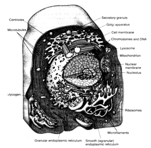
The cell is not merely a bag of fluid, enzymes and chemicals, it also contains highly organized physical structures, may of which are called organelles, and the physical nature of each of these is equally as important to the function of cell as the cell's chemical constituents. For instance without one of the organelles, the mitochondria, more than 95 percent of the energy supply of cell would cease immediately. Some principal organelles of structure of cell are illustrated in figure 7.[127] Including the cell membrane, nuclear membrane, endoplasmic reticulum, Golgi apparatus, mitochondria, lysosomes and centrioles. |
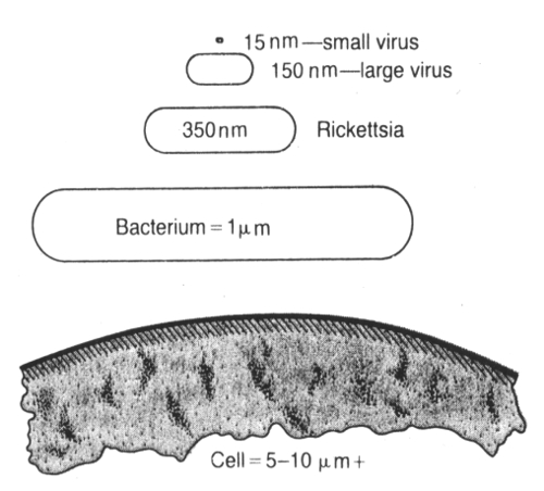
Many of us think of the cell as the lowest level of life. However the cell is a very complicated organism, which requires many million years to develop after the earliest form of life, an organism similar to the present day virus first appeared on earth. Fig. 8.[128] Illustrate the relative sizes of the smallest known virus, a large virus, a rickettsia, a bacterium and a nucleated cell. Showing that the cell has a diameter about 1000 times that of the smallest virus and, therefore, a volume about one billion times that of the smallest virus. Corresponding, the functions and the anatomical organization of the cell are also far more complex than those of virus without nucleus. The essential life giving constituents of very small virus is a nucleic acid embeded in a coat of protein but without nucleus. This nucleic acid is composed of the same basic constituents (DNA and RNA) as found in mammalian cells, and it is capable of reproducing itself if appropriate conditions are available. Thus, the virus is capable of propagating its linkage from generation to generation and, therefore, is a living structure in the same way that the cell and the human beings the living structures. As life evolved other chemical besides nucleic acid and specialized function begin to develop in different parts of the virus. A membrane formed around the various, and inside the membrane a matrix appeared, specialized chemicals develop inside the matrix to perform special functions, many protein enzymes appeared, which are capable of catalyzing chemical reactions, and therefore, of determining the organism's activities. In still later stages particularly in the rickettsial and bacterial stages, organelles developed inside the organism, representing physical structures of chemical aggregates that perform functions in a more efficient manner that can be achieved by dispersed chemicals throughout the fluid matrix. Finally, in the nucleated cell, still more complex organelles developed, the most important of which is the nucleus itself. The nucleus distinguishes this type of cell from all lower forms of life, this structure provides a control centre for all cellular activities, and it provides for exact reproduction of new cells generation after generation, each new cell having essentially the same structure as its proginitor. |
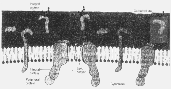
Essentially all organelles of the cell are liked by membranes composed primarily of lipids and proteins. These membranes include the cell membrane, the nuclear membrane, the membrane of the endoplasmic reticulum, and the membranes of mitochondria, lysosomes, golgi apparatus as well as still others. The lipids of the membranes provide a barrier that prevents free movement of water and water soluble substances from one cell compartment to other. The protein molecules in the membrane, on the other hand, often penetrate all the way through the membrane. Also many of the membrane proteins are enzymes that catalyze a multitude of different chemical reactions.
The cell membrane which completely envelopes the cell, is a very thin, elastic structure only 7.5 to 10 nanometers thick. It is composed of proteins and lipids. The approximate composition is:
Proteins = 55%
Phospholipids = 25%
Cholesterol = 13%
Other lipids = 4%
Carbohydrates = 3%
Fig.9: |
The cytoplasm is filled with both minute and large dispersed particles and organelles ranging in size from a few nomometers to many micrometers. The clear fluid portion of cytoplasm in which the particles are dispersed is called cytosol. This contains many dissolved proteins, electrolytes, glucose and minute quantities of lipid compounds. The portion of cytoplasm immediately beneath the cell membrane frequently contains interwoven net of microfilaments composed mainly of actin fibrillae. These provide a semi solid gel-like support for the cell membrane. This zone of cytoplasm is called the etcoplasm. Dispersed int he cytoplasm are neutral fat globules, glycogen graunules, ribosomes, secretory granules, and five especially important organelles, the endoplasmic reticulum, the golgi apparatus the mitochondria, the lysosomes and peroxisomes. |
(v) RibosomesFigure 7 illustrates in the cytoplasm a network of tubular and flat vesicular structures called the endoplasmic reticulum. The tubules and vesicles all interconnect with each other. Also their walls are constructed of lipid bilayer membranes containing large amount of proteins, similar to the cell membrane.
(vi) Golgi ApparatusAttached to the outer surface of many parts of the endoplasmic reticulum are large numbers of small granular particles called ribosomes, where these are present. The ribosomes are composed of a mixture of ribonucleic acid (RNA) and proteins and they function in the synthesis of protein in the cells.
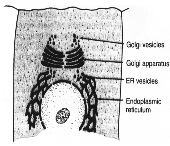
The golgi apparatus, illustrated in fig. 10[129] is enclosed related to the endoplasmic reticulum. It has membranes similar to those of the angular endoplasmic reticulum. It is usually composed of four or more stacked layers of thin, flat enclosed vesicles lying near the nucleus. The golgi apparatus functions in association with the endoplasmic reticulum. As illustrated in the above figure 10, small "transport vesicles" also called endoplasmic reticulum vesicles or simply ER vesicles, continually pinch off from the endoplasmic reticulum and shortly thereafter fuse with the golgi appratus. In this way, substances are transported from the endoplasmic reticulum to golgi apparatus. The transported substances are then processed in golgi apparatus to form lysosomes, secretary vesicles or other cytoplasmic components. |
(viii) MitochondriaLysosomes are vesicular organellers formed by the golgi apparatus that then become dispersed througout the cytoplasm. The lysosomes provide an intracellular digestive system that allows the cell to digest and thereby remove the unwanted substances and structures, especially the damaged or foreign structures such as bacteria. The lysosomes illustrated in fig. 7.
(ix) NucleusThe mitochondria are called the "power houses" of the cell, without them, cells would be unable to extract significant amounts of energy from the nutrients and oxygen, and as a consequence essentially all cellular functions would cease. As illustrated in the cell of figure 7, these organelles are present in essentially all proportions of the cytoplasm. Mitochondria are concentrated in those portions of cell that are responsible for the major share of its energy metabolism.
Mitochondria are self replicate, which means that one mitochondrion can form a second one, a third one and so on, whenever there is need in the cell for increased amounts of ATP. In deed the mitochondria contain de-oxy-ribo-nucleic acid (DNA). Similar to that found in the nucleus. DNA is the basic substance of the nucleus that controls replication of the cell. This substance plays a similar role in the mitochondrion replication process. Many proteins and lipids that have already been formed in the cytoplasm are incorporated into the mitochondria as they enlarge and bud off to form new mitochondria.[130]
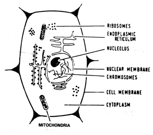
The nucleus is the control center of the cell. Briefly the nucleus contains large quantities of DNA which we have called genes for many years. The genes determine the characteristics of the protein enzymes of the cytoplasm and in this way control cytoplasmic activities. They also control reproduction, the genes first reproduce themselves and after this the cell splits by a special process called mitosis to form two daughter cells, each of which receives one of the two sets of genes. Fig. 11 (a) illustrates the light microscopic appearance of the interphase nucleus (period between mitosis) showing darkly staining chromatin material throughout the nucleoplasm. During mitosis, the chromotin material becomes readily identifiable as the highly structured chromosomes. |
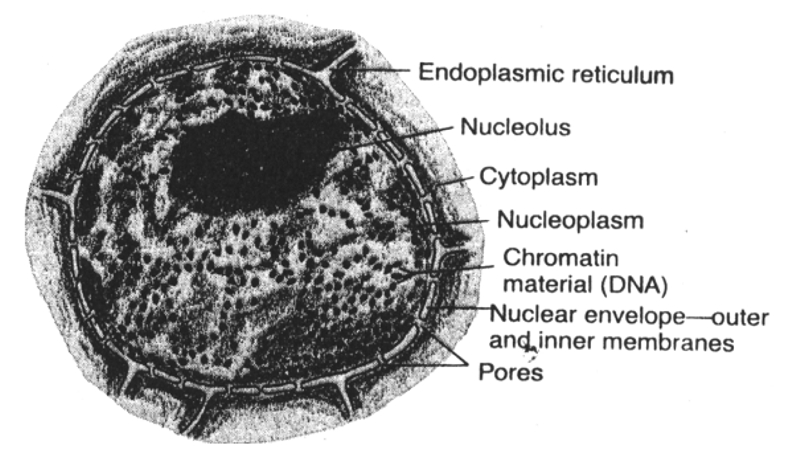
The nuclear envelop is frequently called the nuclear membrane. However, it is actually two separate membranes, one inside the other. The outer membrane is continuous with the endoplasmic reticulum, and the space between the two nuclear membrane is also continuous with the compartment inside the endoplasmic reticulum.
 Doctoral Thesis, JVBU