 Jethalal S. Zaveri
Basic Functions of the System
Blood is 'the essence of life', for this vital fluid carries to every individual cell in the body the nutrients necessary for producing energy and the raw materials necessary for tissue growth, maintenance and repair. It is vital too in clearing away all the waste products of the cells' various activities. It also acts as the body's policeman, liquidating any potentially dangerous invaders. Chemical messengers and other vital substances, too, need a transporting medium to distribute them through the body to keep the cells alive and active. The blood circulatory system with its intricately branching and inter-connecting tubes provide these services. Thus, the basic functions of this system are in the area of transport—carrying anything and everything that needs to be shifted from one part of the body to another. Oxygen from the air that is breathed into the lungs and nutrients absorbed from the intestines are carried and distributed to all the body cells. Hormones—chemical messengers—that regulate and co-ordinate the activities of the body, are carried by the blood, from the glands that secrete them, to the target cells on which they act. It also provides a continual supply of water for the needs of the body. The waste-products such as carbon dioxide (C02), urea, etc. removed by it are carried to the lungs, kidneys and liver, where they are processed and excreted. Finally it is an important link in the body's system of temperature-regulation.
Organs of the System
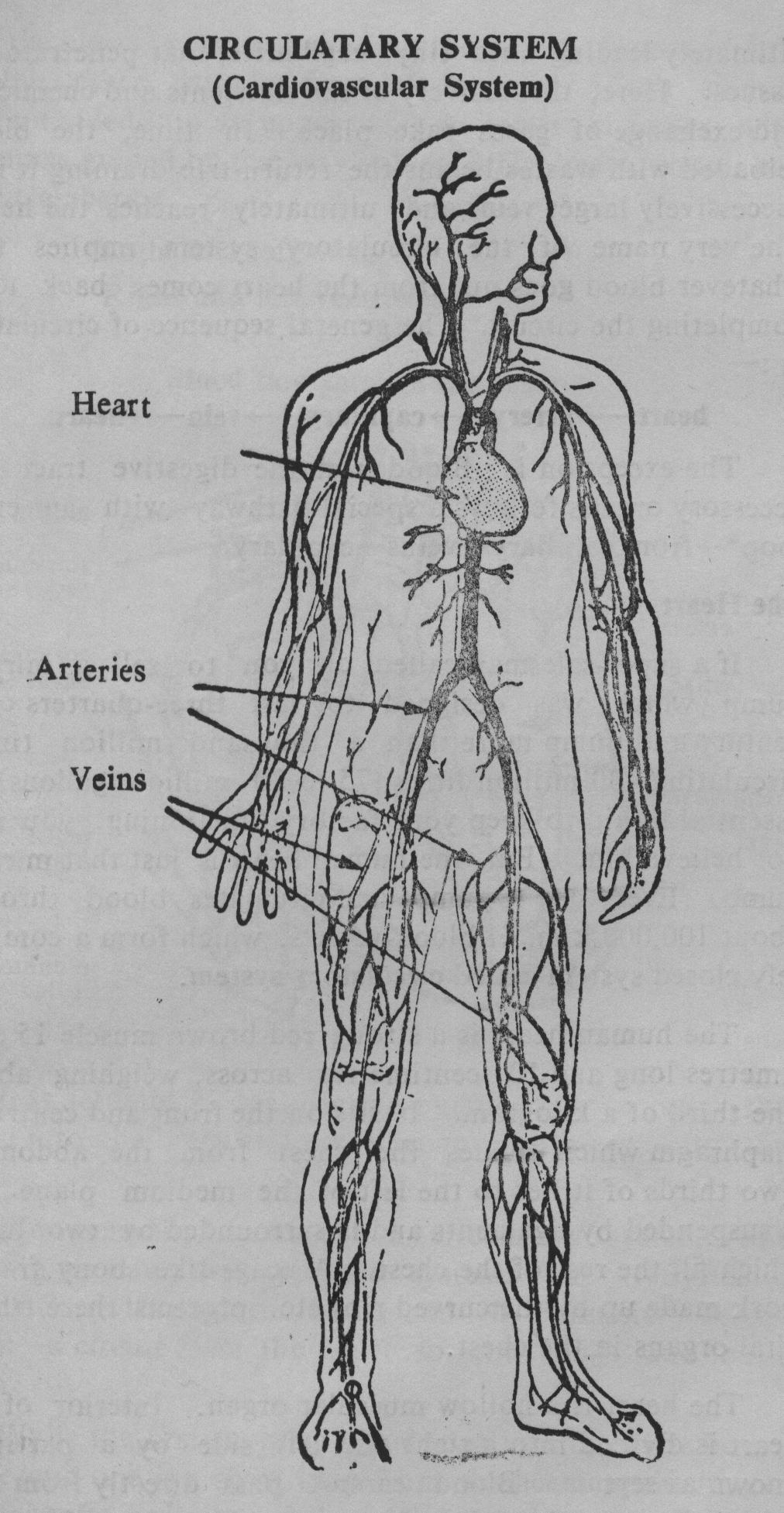
The propulsive force that keeps the blood moving is the steady beating of a powerful pump—the heart. The contractions of the heart drive blood into arteries which branch and rebranch into smaller tubes, arterioles ultimately leading into tiny capillaries, that penetrate the tissues. Here, the delivery of the nutrients and chemicals, and exchange of gases take place. In time, the blood reloaded with wastes begins the return-trip draining it into successively larger veins and ultimately reaches the heart. The very name of the circulatory system implies that whatever blood goes out from the heart comes back to it, completing the circuit. The general sequence of circulation is:—
heart→artery→capillary→vein→heart.
The exception is: Blood from the digestive tract and accessory organs follows a special pathway with an extra loop[*]→from capillary→veins→capillary.
The Heart
If a super-salesman called on you to sell a miracle pump which was designed to last three-quarters of a century and pump more than a thousand million times, circulating 400 million litres (75 to 80 million gallons) of essential liquid to keep your factory functioning, you may not believe him. But the human heart is just that miracle pump. Every day it pumps and circulates blood through about 100,000 k.m. of blood-vessels, which form a completely closed system called circulatory system.
The human heart is a strong red-brown muscle 15 centimetres long and 10 centimetres across, weighing about one-third of a kilogram. It sits on the front and centre of diaphragm which divides the chest from the abdomen. Two thirds of it lies to the left of the medium plane. It is suspended by ligaments and is surrounded by two lungs which fill the rest of the chest. A cage-like bony framework made up by the curved ribs etc. protects these three vital organs in the chest.
The heart is a hollow muscular organ. Interior of the heart is divided into a right and left side by a partition known as septum. Blood cannot pass directly from the left to the right side of the heart or vice versa. However an congenital defect—'a hole in the heart' is quite common and the blood is shunted through the defect, from one side to the other (usually from left to right). Each side is again divided into an upper and a lower chamber with openings guided by one way valves. The heart, thus, has four clambers:
Right and left atria
Right and left ventricles-
Blood flow through the heart
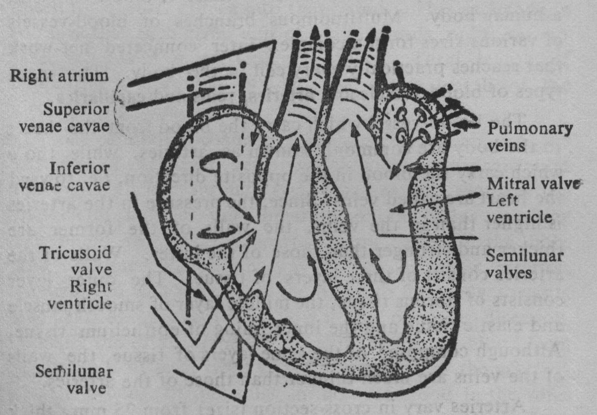
It is actually a dual action-pump—one to send the blood into the lungs and the other to push it out into the body.
We generally speak of a circulatory system, but actually we have two separate systems viz. (i) Systemic Circulation—through the body, and (ii) Pulmonary Circulation—a circuit from the heart to the lungs and back again.
While the first supplies blood to all parts of the body, the second one is a short loop from the heart to the lungs. Both circuits begin and end in the heart, and there is no mixing between them.
The Functioning of the Heart
One sometimes wonders how the heart manages to pump day and night for seventy years or more without resting. The answer, of course, is that the heart does rest, between every beat. The resting period is called diastole and, its duration is twice as long as that of systole, which is the period of muscular contraction.
Blood-vessels—Arteries, Veins and Capillaries
There are more Than 100,000 kms. of blood vessels in a human body. Multitudinous branches of blood-vessels of various sizes form a complex inter connected net-work that reaches practically every cell in the body. The main types of blood-vessels are arteries, veins and capillaries.
The blood-vessel which carry the blood from the heart to the body are commonly known as arteries, while those which carry the blood in the opposite direction, i.e. toward the heart are called veins. Since, the pressure in the arteries is higher than in the veins, the walls of the former are thicker and stronger than those of the latter. Walls of the arteries consist of three layers of tissue. The outer layer consists of fibrous tissue, the middle layer of smooth muscle and elastic tissue and the inner lining of epithelium tissue.-Although composed of the same layers of tissue, the walls of the veins are much thinner than those of the arteries.
Arteries vary in cross-section (size) from 25 mm. thick aorta down to less than 1.5 mm. in diameter. The bloodflow is helped along by rhythmic contractions in the muscular artery-walls.[**] Arteries are generally embedded deep into the muscles as a protection from all but severe injuries.
Oxygenated blood leaves the heart chamber via the aorta which emerges from the left ventricle and soon branches into two, one going upwards to the heart and the other downwards to the trunk and limbs. Aorta divides into arteries which again divide into smaller vessels called arterioles. The division progresses and ends into tiny thin-walled capillaries. These gossamer cobwebs link the smallest arterioles to the smallest venules. The wall of a capillary is composed of a single thin layer of endothelial cells which permits the passage of water and other small molecular substances. Their diameter is approximately that of a red blood cell.
Capillaries are so small (.006 mm. dia.) that the red blood cells have to pass through them in single file. They permeate the tissues and service the body-cells directly. Oxygen, food materials and hormones transported in the blood are transferred to the cells, while cell-products, produced for export, diffuse into the blood. This exchange is so rapid that each given unit of blood spends only one to three seconds in a particular capillary. Total length of capillaries is more than 99% of the entire length of the circulatory system.
The capillaries coalesce into venules, which are the branches of the larger veins, which eventually drain into the right auricle through two venae cavae. Contraction of skeletal muscles puts pressure on the veins pushing the blood towards the heart. Many veins of the limbs and abdomen have one-way valves in their interior, so arranged that they allow the blood to flow only towards the heart and prevent it to flow in the opposite direction. Usually the total diameter of veins returning blood from an organ is at least twice the total diameter of the arteries. This is because of the sluggish flow of blood in the veins which requires the greater capacity to maintain the balance between the incoming and the outgoing blood volume. All the veins of the systemic circulatory system (except for the cardiac veins) empty into either the superior or inferior vena cava which drains into the right auricle.
Cardiac Cycle
A full cardiac cycle takes less than a second. Yet during that brief time a complicated sequence of events takes place. The working part of the heart is its muscular walls. They contract and relax rhythmically. This rhythm is self-initiated requiring no nervous or chemical stimulation. During relaxation, blood from the tissues with a high carbon-dioxide content carried by veins, enters the upper right chamber (right auricle). At the same time, oxygenated blood from the lungs carried by the pulmonary vein fills up the upper left chamber (left auricle). Then the walls contract. The pressure inside the chamber increases. The blood is forced out from both the auricles The impure blood from the right auricle rushes first into the right ventricle through the one-way valve and then out of the latter into the pulmonary artery from where it reaches the lungs. Simultaneously, the oxygenated blood from the left auricle rushes first into the left ventricle and then out of the latter into the aorta, from where it is distributed throughout the body through arteries and ultimately into tiny capillaries.
Red blood-cells carrying oxygen squeeze through the tiny capillaries in a single file and deliver the load of oxygen to the cells and get reloaded with carbon-dioxide- The flow now reverses its direction and travels towards the heart through the veins and ultimately drains into the upper right chamber by way of two venae cavae. By the time the blood is ready to move towards the heart, the pressure has dropped down to almost zero and the heart is not capable of exerting suction pressure. The flow towards the heart must, therefore, be aided by something outside the system. Such assistance is given by the contraction of muscles which squeeze the veins and push the blood upward while one-way valves prevent the back-flow.[1]
The phase of contraction is called systole, while that of relaxation is called diastole. When the heart rate is normal i.e. at the rate of 70-72 beats per minute, diastole lasts for.49 seconds and systole for.36 seconds. During heavy physical exertion the heart-rate increases to 170 beats per minute. At this rate, diastole is only.12 seconds while systole lasts for.23 seconds.
It should always be remembered that with every heartbeat, first both auricles and then both ventricles contract sending blood out; then both sides relax and receive blood from both routes.
Nourishment of the Heart
For proper functioning, the heart, like any other organ, must be adequately nourished. In fact, it needs ten times the nourishment required by the other organs and tissues. The nourishment is not, however, obtained from the blood passing through its chambers, but from its own blood supply. It is fed by two (right and left) coronary arteries which arise from the aorta just after its emergence from the left ventricle. Both branch out again and again surrounding the heart-muscle and nourish it adequately. Heart-attacks or coronaries are caused by narrowing or partial blockade of these arteries.
Cardiac Output
Under resting conditions each systole pumps about 70 ml. of blood into the aorta. This is called stroke volume. The cardiac output is defined as—stroke volume x heart-rate.
With an average heart-rate of 70-72 beats per minute, the cardiac output comes to about five litres.[2] Thus in the space of just one minute, a volume equivalent to all the blood in the body passes through the heart- But during heavy exercise, the amount pumped can increase to over 25 litres per minute.
The cardiac output does not remain constant but is adjusted to meet the varying needs of the body. The heart is capable of increasing its output by fivefold or even more. It has been estimated that during a man's life-time a total of 500,000,000 litres blood is pumped. Under normal conditions the liver receives 26% of the total cardiac output; the kidneys 24%, the skeletal muscles 15%, the brain 14% and the heart 5%.
Heart-rate steadily slows down as one gets older; it is 140 before birth, 90 as a child and 70-72 as an adult.[3]
The Blood-Stream
Blood is a red-coloured fluid with a salty taste and a characteristic odour. Its specific gravity is only slightly more than water, about 1.055, but its viscosity is about five times as high and hence it is not as free-flowing as water. The volume of blood varies with the size and the age of a person. An average sized adult man has about 5 litres and an adult woman 4.5 litres of blood circulating in the system.
Besides the oxygen, a variety of other substances must be delivered to the cells and tissues to keep them alive and vigorous. Glucose, fat, amino-acids and water are some of the more common, while copper and cobalt, iodine and phosphorus are a few of the uncommon requirements of the cells.[4] Salts, minerals, vitamins and hormones are also carried by the bloodstream for delivery to appropriate places. At the same time, carbon-dioxide and other waste products are also carried by the blood to the kidneys and other sites for excretion.
Thus blood is ever-changing, yet its overall composition remains surprisingly constant.
Composition of the Blood
Blood is composed of about 55% fluid and about 45% solid matter. A single drop of blood contains more than 250 million blood cells, suspended in a clear fluid. The apparently homogeneous fluid is actually teeming with countless numbers of substances of thousands of different kinds each with its own specific job to do. The main components of the blood are:
Blood plasma—a clear straw-coloured fluid in which are dissolved salts, proteins, fats, sugar, hormones, vitamins, as well as waste products such as urea and lactic acid. Indeed, this clear, rather sticky, fluid is far more complex mixture - perhaps one of the most complex mixtures in the universe. 90 to 92 percent of the volume of plasma is water and 8 to 10 percent other constituents. Water, not only serves as the fluid medium of transport, but also acts as a solvent that brings together an intricate variety of chemical substances and helps them to react.
The solid elements also called formed elements, suspended in the fluid plasma, make up 45 percent of the total volume of blood. They include three main types: red blood cells (RBC) or erythrocytes, white blood cells (WBC) or leukocytes and platelets or thrombocytes.
Normal blood contains approximately:
Red cells 5 x 10¹² per litre
White cells 8 x 109 " "
Platelets 250 x 109 " "
Red Blood Cells (Erythrocytes)—these are small flattened bi-concave discs. 3000 of them would be needed to measure an inch. It contains a red pigment, haemoglobin and the huge masses of red cells, hundreds of millions in each drop that together give blood its rich red colour. The shape and structure of these cells represent an extreme of adaptation for its function. They are the gas carriers of the body.
They should be correctly called corpuscles rather than cells, because they have no nucleus but they are often called red cells.
There are five million red cells per cubic millimetre. Every second three million cells die and equal number of them are born. In the embryo, red cells are formed in the liver, spleen and bone-narrow. At some time before birth, the liver and spleen shut down their red cells production, and the function is taken over entirely by the bone-marrow. A red cell has an average life-span of 120 days during which it makes 300,000 trips, travels more than 1000 km., carrying three quadrillion molecules of oxygen.[5]
Haemoglobin has a strong affinity for oxygen and when they come into contact the oxygen is taken up, forming oxyhaemoglobin. This process normally takes place in the lungs. Oxygenated arterial blood has a bright red colour while that in the veins, having lost its oxygen, has a bluish-purple hue.
Haemoglobin of the red cells is an iron containing pigment called haem, combined with a protein, globin. Total amount of iron in the haemoglobin of all the red cells is about 3 grams. But it is priceless, as one cannot live without it. A continual intake of iron in diet is essential for the promotion of new RBC. Men lose only about 1 mg of iron each day but a woman loses about 20 mg during each normal menstrual period, A minimum of 5 to 15 mg of iron must be consumed in the daily food. Need for iron increases greatly during pregnancy. Each haemoglobin has four iron atoms which combine loosely with four molecules of oxygen in the lungs. There are about 280 million molecules packed into each of the 5 million red Wood cells in each cubic millimetre of blood. Contents of haemoglobin in the blood is an index of health.[6]
White Blood Cells (Leukocytes). These are essentially colourless, as they do not contain haemoglobin. They are larger than red cells. They are not uniform in size, shape and appearance. Unlike red cells which are carried passively along in the blood-flow, white cells move actively. The main functions of most of the white cells are performed outside the actual blood, in the neighbourhood of tissues. These soldiers of the body are second-line defenders, (skin and mucous membranes are the first line), against foreign invaders ready to fight to death, if need be. Red cells outnumber the white cells by about 700 to 1. The life-span of a white cell is extremely variable. It may be only a few hours, or with luck may be measured in months. On the whole, the life of the white cell is harder than that of the red cell. It is 3ikely to meet an untimely death in the service of the body. A continuous production of new white cells is, therefore, essential. The red bone-marrow is also the site of the formation of the leukocytes. Some others are produced in the various lymph nodes, the spleen, the thymus gland and the tonsils.
Platelets (Thrombocytes)
They are also called clotting cells (thrombo means clot). They are more numerous than white cells, but less so than red cells. They live in the blood for five to eight 4ays after which they are destroyed in the spleen and other organs. They are a key link in the mechanisms for the prevention of blood loss and they go into action when a blood-vessel is cut or pierced and blood is being lost. A substance called platelet factor initiates a chain of chemical reactions leading to the formation of a clot which effectively plugs the cut or hole. Blood-clotting is a complicated chain of chemical reactions involving 13 separate factors.
Blood-Pressure
Blood-pressure is the propelling force in the arteries. It is a measure of the pressure exerted on the walls of the arteries[7] by the flow of blood.
It depends upon—(a) the output of the heart, and (b) the resistance encountered by the flow in the smaller arteries and arterioles. The pressure in arteries and arterioles reaches a peak called the systolic pressure with each contraction of the heart and then gradually decreases to a minimum, the diastolic pressure, before the next contraction. It is commonly measured in the artery just above the elbow and is always expressed as two figures e.g. 120/80 in healthy young adults. When outside these limits, it is either high or low.
Blood-pressure is an extremely fluctuating physiological function and it is rather difficult to define 'normal' blood-pressure. Firstly, it rises and falls at different times of the day. Secondly, it is higher when one is actively exercising than when one is resting or sleeping. Again, when one is emotionally upset, it rises, and falls down when one becomes calm and quiet. And so the pressure is arbitrarily defined as 'normal' where more than 90 percent of the blood-pressure of population happens to be.
If the pressure is too low, the tissues may not receive adequate supply of nutrients. A few years ago, low pressure was considered undesirable. Nowadays, however, if there are no adverse symptoms, such as dizziness, fatigue or fainting, the low pressure is regarded a protection from the development of atherosclerosis or hardening of the arteries.
Constant high blood-pressure, on the other hand, is definitely injurious. Hardening of the arteries (atherosclerosis is caused by the deposition of the blood clots, fats and calcium on the inside walls of the arteries, making the normally soft elastic arteries to become hard, inelastic and partly or completely blocked'.
The risk of developing atherosclerosis is directly related to the level of blood-pressure. If the arteries to the heart, called coronaries, are blocked, death of heart cells is inevitable and a heart-attack may follow. If the arteries to the brain become obstructed, strokes may occur. Thus continuous high blood-pressure or hypertension is the indirect cause of death viz. heart-attack and stroke.
Emotional stresses, like excitement, agitation or annoyance also cause hypertension. Whenever one encounters a stressful situation, an innate mechanism is automatically put into action, causing an excessive flow of adrenalin and resulting in a marked rise in a blood-pressure, acceleration of heart-rate and other physiological changes. If the mechanism is repeatedly activated, e.g. due to stressful situations built into our daily life, it will ultimately result in hypertension. Fortunately, there is also another innate protective mechanism which produces diametrically opposite conditions. Regular practice of relaxation and meditation can activate the protective mechanism which normalizes the blood-pressure.
Unlike the heart-rate, the blood-pressure tends to rise as one grows older. For a new-born baby it is 40; it rises quickly to 80 by the end of the first month, and then continues to rise more slowly. By the tenth year, it is about 100. At puberty, the adult level of 100-120 is reached. After 25 years, it creeps up by about.5 per year. It is 140 at 60 and 160 at 80.
THE LYMPHATIC SYSTEM
The lymphatic system communicates with the blood circulatory system and is closely associated with it. It has several functions which complement the function of the blood circulatory system
Though technically the blood circulatory system is a closed system, there is a continuous leakage of fluid and proteins out of the capillaries into the tissues. To prevent the blood from thickening, the fluid must be returned to the system. This service is provided for by the 'lymphatic system'. Almost all tissues have lymphatic channels, which drain excess fluid into this system. It can be regarded as a part of the circulatory system. Besides returning body-fluid to the blood, it transports substances such as fats, hormones and enzymes from their manufacturing sites to the blood-stream.
Lymph—the fluid which has passed into the lymphatic vessels—is a watery plasma-like fluid. Wherever there are blood-vessels, there are also lymph-vessels. Lymphatic system resembles blood-system. Lymph capillaries unite to form large vessels which unite, in turn, to form larger vessels. Finally, they converge into two main ducts which empty into the various portions of the blood-system.
Lymph-capillaries are very thin-walled tubes which are even more permeable than blood-capillaries, but it is a one way permeability. Once, fluid, protein and other particles have entered, they stay inside and cannot leak into the tissue.
Lymphatic system is also the head quarters for the important line of defence against diseases. It provides the body with lymphocytes and other anti-body-producing cells for defending it against disease-germs and other foreign invaders.
SPLEEN
Strange as it may seem, spleen is classified as a lymphoid organ. It is a dark purple coloured organ, 14 cms. long, 8 cms. broad and 3 to 4 cms. thick. It is situated directly below the diaphragm above the left kidney, and behind the stomach. It is a versatile organ performing a number of different functions. Some of the important functions are:—
- The spleen acts as a filter capturing and removing bacteria, debris, parasites and other infectious agents from the blood-stream.
- It is the grave-yard of the red blood cells. Old and worn-out cells rupture as they try to squeeze through the narrow channels of the spleen and are destroyed. The haemoglobin from the destroyed cells is broken down; iron and globin are salvaged and returned to the blood-stream for recycling.
- Spleen joins with other lymphoid organs (particularly the thymus) in the immune response of the body. They produce antibodies (immunoglobulins) which are released into the blood-stream conferring humoral immunity against specific bacterial infections.
- Before birth, spleen manufactured red blood-cells. It is ready to regain its ability to produce red cells in an emergency such as anemia.
- It acts as a blood-lake and a reservoir for red cells sending stored blood into circulation when needed. The blood stored in the spleen has a higher concentration of blood-cells and can increase the hematocrit 8 of the system by as much as 3 to 4%.
When you take a pulse at the wrist, you are feeling the contractions of arteries and not those of heart.
In its return-journey through veins, blood from the limbs and abdomen must flow against gravity. Three important mechanisms aid in the venous return: (i) vasomotor action, (ii) muscular pump, and (iii) respiratory pump.
Actually the normal range extends from 60 to 100.Incidentally heart-rate of a mouse is 700, that of a rabbit 150, while an elephant's is only 25 beats per minute.
The normal level of haemoglobin in the blood is 13.0 to 18.0 3rams/100 ml for men and 12.0 to 16.0 grams/100 ml for women.