 Jethalal S. Zaveri
The nervous system is the most highly developed communication system in the human body. It co-ordinates and controls the work of other systems (of the body) and through them controls the functions of the body as a whole. It makes possible a range of adaptive responses to changes in the environment. Such responses are central to the behaviour of all living organisms and are known as homeostatic responses. If the body were unable to adapt to extremes of heat or cold, the healthy functioning of the vital organs would be threatened. Recognition of stimuli, storage of information (memory), communication between the various parts of the body, and the extension of effective responses are functions of this system. That is why it is considered to be one of the two most important systems of the body. Its failure will result in total cessation of its activities, paralysis of all the organs and ultimate stoppage of minute vital processes. One would be unable to use one's muscles, unable to move one's hands, blink one's eyes, to sit or stand, even to breathe. The nervous system is intimately associated with the endocrine system, and both together co-ordinate the body's activities and integrate the organism.
The nervous system is made up of two parts: an outer or somatic system and an inner or visceral system. The former consists of the sense-organs, the muscles, bones and joints. The latter controls the internal organs such as the heart, glands, blood-vessels and intestines. The activities of both are co-ordinated by the Central Nervous System (CNS).
Structural Organization
The structural arrangement of the nervous system is the basis for its division into two parts:
- Central Nervous System (C.N.S.) consisting of the brain and the spinal cord.
- Peripheral Nervous System (P.N.S.) consisting of 31 pairs of spinal nerves, 12 pairs of cranial nerves.
The functional relationships are the basis for differentiation of another division, the autonomic nervous system.
The Central Nervous System
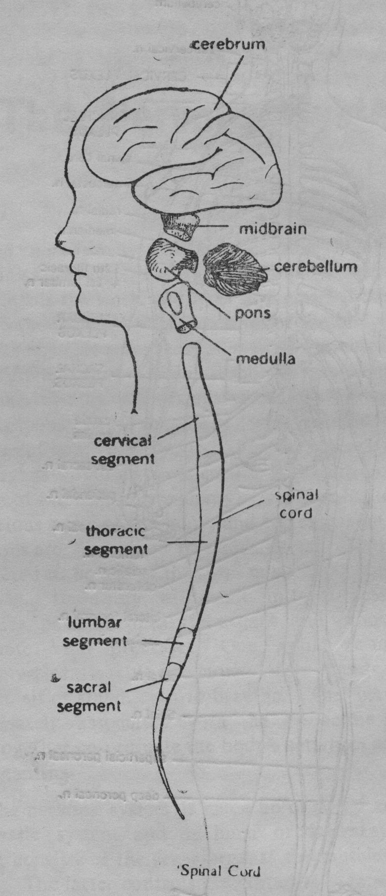
The brain and spinal cord merge smoothly into one another without any obvious demarcation; the portion above the foramen magnum (the opening in the base of the skull) is considered as the brain while the portion below it is the spinal cord. The brain directs all conscious movements, and a number of unconscious, in voluntary actions as well.
The spinal cord, a shining rope of nerve cells, runs along the channel formed by the vertebral column.
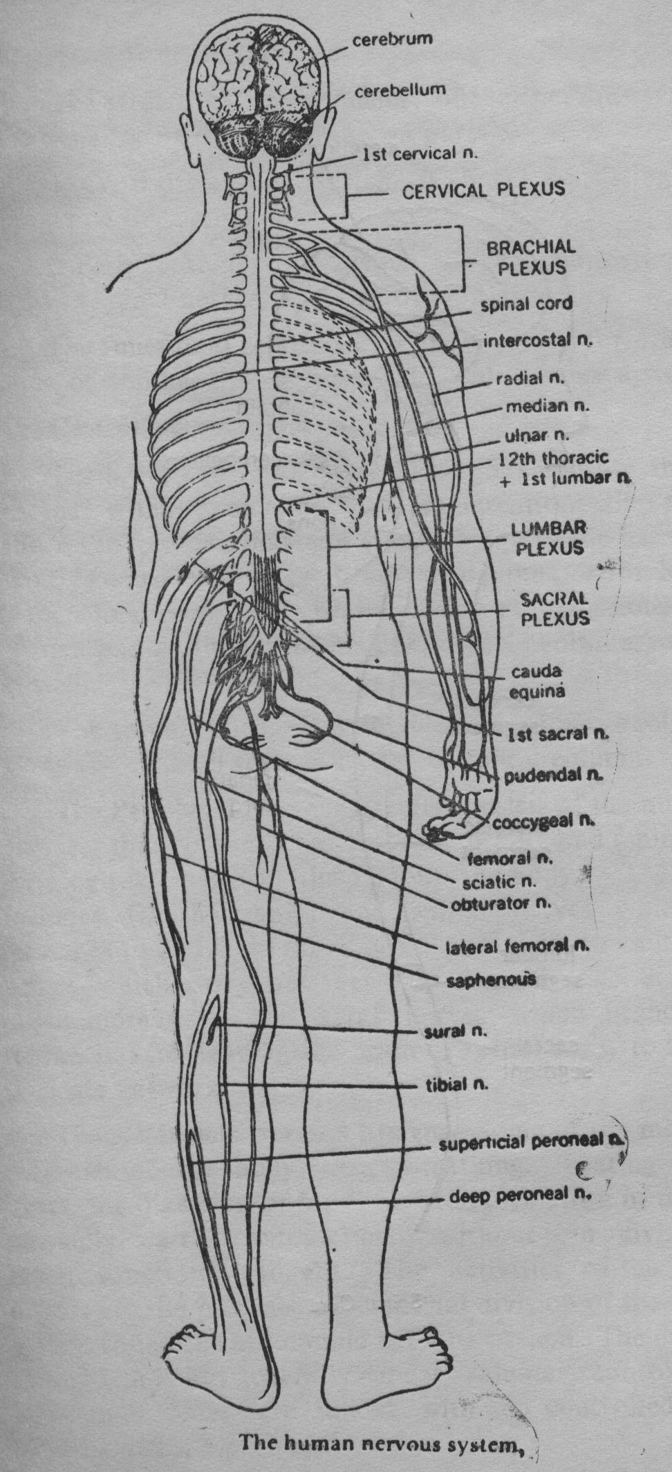
The Peripheral Nervous System consists of the numerous nerves, that branch out from the brain and spinal cord forming the network throughout the body. The P.N.S. includes (i) the cranial and spinal nerves which carry messages to and from the C.N.S. and (ii) the autonomic nerves, which carry messages only from the C.N.S. There are hundreds of peripheral nerves which branch and rebranch, after leaving the brain or spinal cord, to form an intricate network.
The Autonomic Nervous System—Some of the most vital activities of the body, viz., breathing, beating of the heart, the digestion of food, and the formation of urine are normally carried automatically without our giving them much conscious thought. The activities of the internal organs are the province of a special division of the nervous system called the autonomic nervous system. The action of this system is not generally under voluntary control. The autonomic fibres are linked with and controlled by the hypothalamus, a part of the brain.
There are two main divisions of this system—(i) The parasympathetic and (ii) sympathetic. The action of the two divisions is antagonistic. In general, the parasympathetic system exerts its influence during the time of rest; the sympathetic system prepares the body for emergency conditions and aids in the mobilization of body resources and energy production.
Functional Organization
More than 10 billion neurons comprise the human nervous system. Through the intricate network run impulses that are the foundations of creative thoughts as well as desires. Innumerable bits of data continually flood the body's sensory-organs and pass from the receptors along the peripheral system to the central nervous system. The latter system screens and evaluates the incoming impulses; stores important data for future retrieval; formulates decisions and initiates actions via impulses sent along the peripheral nerves to the muscular system.
Thus the nervous system has two basic functions—(i) the detection and processing of information from within and outside the body and (ii) production and regulation of movement by muscle-section. Some parts of the brain are also responsible for the control of emotions and the storage of information and are also concerned with personality and intellect.
We shall now examine the components of each division of the nervous system in greater detail.
1. The Central Nervous System
A. BRAIN
The brain is the most complex and the most important part of the nervous system. In it are the sites of consciousness, thought, memory, creativity, speech, vision, hearing, smell, control of endocrine glandular secretions and autonomic (involuntary) functions, and the will to carry out purposeful actions. It is only because of the brain (his or hers and our own) that we know anything of the personality of an individual. Inspite of incredibly immense progress made by the electronics technology, a living human brain is a far more compact, yet complex and efficient apparatus than the best artificial intelligence modern technology could devise.
It is the seat of personality of love and hate, pleasure and pain—the centre of awareness of the world around us—the source of intellect and creativity. The brain is that portion of the central nervous system which is safely enclosed within the hard bony helmet i.e. the cavity of the skull.
The adult human brain weight about 1.4 kgs. It is composed of some 30,000 million neurons and five to ten times that number of glial cells.
The brain receives about 17% of the cardiac output and about 20% of the total oxygen consumed by the body even though it is only about 2% of the body weight. It has three major divisions:
- The forebrain consisting of the two cerebral hemispheres (the cerebrum) and interbrain (diencephalon).
- The midbrain (mesencephalon)
- The hindbrain comprising of
- the pons,
- the medulla oblongata,
- the cerebellum.
The midbrain, pons and medulla oblongata together constitute the brainstem.
Cerebrum
The cerebrum is the most prominent portion of forebrain.
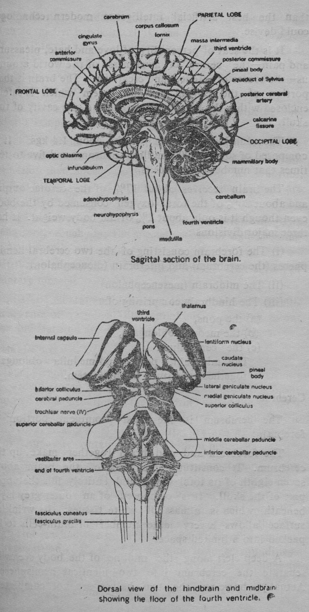
The forebrain and mid brain together make up the cerebrum. It constitutes the bulk of the brain about seven-eighth of its total weight and entirely fills the upper part of the skull. It is composed of an outer grey layer beneath which is a mass of white tissues. Its wrinkled surface allows a very large number of brain-cells to be packed into a limited space.
A deep cleft along the midline of the body seems to separate the cerebrum into two identical hemispheres. Actually, however, the two halves are not completely separated: deep inside, a thick bank of nerve fibres as well as three smaller bands interlink the two halves and provide for an interchange of information between them. Each hemisphere controls the voluntary movements of the other side of the body. Each hemisphere is further divided into four main regions called lobes:
(i) Frontal lobe; (ii) Parietal lobe; (iii) Occipital lobe; and (iv) Temporal lobe.
The cerebrum contains millions of neurons that are the basis of mind. In fact, man's ascendancy over his environment and other species is due to the size of his cerebrum, which is bigger in man than in any other animal. There are three varieties of activity associated with cerebrum:
- Sensory perception: The perception of pain, temperature, touch and the special senses of sight, hearing taste and smell.
- The initiation and control of the contraction of voluntary muscles.
- The mental activities involved in memory, intelligence, sense of responsibility, thinking, reasoning, moral sense and learning are attributed to the higher centres.
Cerebral Cortex
Our memories, hopes, plans, attitudes and personality—are all stored within a thin outer layer of the brain composed of grey matter in the cerebrum called the cerebral cortex. It is crumpled and wrinkled. If stretched out flat, it would cover nearly 4000 sq. cms. Its average thickness is 1.6 to 4 mm. It contains 10 to 14 billion neurons.
Research has yielded elaborate functional maps of the cerebral cortex. Out of the several lobes in which the cerebrum is divided, the frontal lobe is concerned with personality, behaviour and movement. The lowest left.frontal part is the centre for speech. The next are the two parietal lobes which are involved in the analysis of sensations and recognition of the body in relation to its surroundings. Occipital lobes, situated on the top of the cerebellum at the back of the head comprise the visual area of the brain. A separate projection on either side of the brain is the temporal lobe which interprets auditory information. Hidden inside the temporal lobe is the area concerned with taste and smell.
The pre-frontal region which lies in front of the motor area is very well-developed in man. It has no primary sensory or motor functions, but is connected with other cortical regions and with some of the deeper structures of the brain. It is involved in the control of emotions and the storage of information, and it is the part of the brain concerned, above all, with personality and intellect.
A general picture of the abilities of the two hemispheres has been developed by various experimental studies. The left brain reads, writes and speaks fluently and does difficult arithmetic. The right half seems rather stupid in comparison for verbal capabilities, but has a keener sense of shape, form etc. as well as a flair for musical rhythm and melody. Specialization of two halves is not an inherent difference in capacity, for if one side is damaged, the other one can be retrained to take over all functions. Dominance of one half over the other is responsible for "handedness".
Memory is of two types—'temporary' and 'long-term' memory. Each memory is stored in redundant fashion on many different parts of the brain and is multiplying repeated. The storage and retrieval of memories seem to involve both electrical and chemical phenomena including the formation of engram [1] (memory trace). Each of the neurons in a particular engram may be interconnected with numerous other memory traces, and from these interconnections the brain builds up its intricate "filing system".
Thalamus and Hypothalamus. At the base of the cerebral hemispheres, buried deep within the brain, are important structures, including the thalamus and the hypothalamus.
Thalamus is situated near the midline. It is a pair of egg-shaped masses of grey matter. It is predominantly a sensory relay station with incoming fibres from the spinal cord and brainstem and outgoing fibres to the cerebral cortex.
All sensations from the periphery of the body (head, limbs and trunk) are conveyed to the thalamus. It is here that very crude, uncritical sensations reach consciousness. Critical interpretation of these occur in sensory area of the cerebral cortex. If it is damaged, one loses the ability to localize sensations precisely.
Hypothalamus is a mass of grey matter situated below the thalamus, at the base of the brain- Its importance seems quite out of proportion to its size. Although it constitutes only about 1/300 of the total mass of the brain, it is a vital link in the physical and emotional life of the body. It has neural connections to the posterior lobe and vascular connections to the anterior lobe of the pituitary gland. Hypothalamus contains numerous specialized control-centres including—
- Cardio-vascular regulation through participation in autonomic responses, regulation of heart-rate, cardiac output, blood-pressure etc. The activities of both components (sympathetic & parasympathetic) are coordinated and controlled.
- Body temperature regulation—regulation of production and loss of heat by stimulating shivering and sweating.
- Regulation of food intake (both hunger and satiating centres) and gastro-intestinal activity.
- Regulation of water balance—it contains receptors which are sensitive to changes of salt concentration in the blood and controls intake or output of water via ADH (anti-diuretic hormone) and thirst centre.
- Control of circadian rhythms which include wakefulness and sleeping, body temperature cycle etc. and which occur with a periodicity of about 24 hours. Hypothalamus exerts overall control over these rhythms.
- Stimulation of sexual activity in concert with the limbic system.
- Emotional feeling and expression. Feelings and physical accompaniments are integrated by the hypothalamus, limbic system and prefrontal cortex. Centres of anger, fear, pleasure and pain are located in hypothalamus and elsewhere.
This part of the brain is obviously implicated in the most vital bodily functions, since an injury to it may cause emotional outburst, over-eating, obesity and reduction of sexual activity.
Cerebellum and Brainstem. The hindbrain comprises of cerebellum and brainstem. The cerebellum, literally meaning 'little brain' sits behind the brainstem below the cerebrum and is concerned with the fine control and regulation of movement and maintaining balance. It looks very much like a small version of the cerebrum. It is divided into two hemispheres and is a mixture of grey and white matter.
The cerebral cortex plays a key-role in the control of muscular contractions that move body parts. But the part of the brain that makes sure that each muscle contracts not too much or too little but just enough to carry out cerebrum's intentions is the cerebellum. Damage to the cerebellum results in the loss of equilibrium and the person's movements will become jerky and uncoordinated.
The mid-brain, pons and medulla together are called the brain-stem from their resemblance to the stem of a fruit.
The brainstem connects the cerebrum with the spinal cord. Its uppermost part'—the midbrain, is tucked up in the cerebrum. It houses a number of movement coordinating centres and is also responsible for sleep and wakefulness. Beneath this is the pons or bridge connecting it with the cerebellum.
Pons is a bridge-like structure, consisting mainly of white matter and serving as a relay station, linking medulla with the higher cortical centres. Finally the medulla oblongata joins the pons to the spinal cord. It is about one inch long portion of the brain-stem. It merges continuously with the spinal cord below and the pons above. Situated here are the centres for the control of key body-functions viz, the cardiac, vaso-constrictor and respiratory centres.
- Cardiac Centre. A number of nervous and endocrine influences work to regulate the rate of the heartbeat. Cardiac centre in the medulla is an important link in this regulatory system. It works through the vagus nerve which joins with the cardiac branches. Impulses from this centre act to slow down the heart-rate. They act antagonistically to the impulses from the cardiac sympathetic nerves which speed up the heart-rate.
- Vasoconstrictor Centre. This regulates the arterial pressure by constricting the smooth muscles of the arterioles. This centre is itself controlled by higher centres, particularly, hypothalamus.
- Respiratory Centre. This controls the rate and depth of respiration. It is sensitive to the concentration of carbon dioxide in the blood. Physical exercise provokes an immediate increase in the rate and depth of respiration. Emotions such as fear also tend to increase the rate as part of body's automatic preparation for fight or flight.
- In addition to these centres, the medulla contains control-centres for reflex action such as hiccupping, coughing, vomiting, sneezing and swallowing.
Reticular Activating System
An intricate cone-shaped network of nerve-cells which runs through the medulla, pons, mid-brain and up into parts of the thalamus and hypothalamus, is the brain's filter called R.A.S. It acts as a sort of central clearing house for the flood of information that bombards the brain. It lets only strong or novel signals pass upto the higher brain for conscious perception.
Though hundreds of thousands of sensory messages are received by the brain every second, only those which are important are perceived, while the rest are ignored. Thus, there is a remarkable distinction between sensation and perception.[*] The process by which the mind converts raw sensations into perception is complicated. While the sensation varies according to the power of stimuli, perception varies by an infinity of factors, some within the body and the others without. The state of emotion, in particular, may have a profound, and at times, decisive effect.
The R.A.S. action permits one to concentrate on a particular thought or activity, disregarding the background noises and other potential distractions. There is also a limit to the capacity of concentration of the brain in the perception of more than one thing at a time. If, for example, one attempts to hear music and read at the same time, he can concentrate on one or the other, but not both.
In addition, a continual dialogue between the cerebral cortex and the R.A-S. keeps the higher brain awake and alert.
Protection of the Brain
The brain is the most carefully protected structure in the body. Not only is it encased in a rigid helmet of bone - the skull—but within the skull it is wrapped in a series of three separate membranes—the meninges. The three membranes are (from the outside inward) the dura mater (hard mother), the arachnoid membrane and the pia mater (tender mother). Extensions of these three membranes similarly encase and protect the spinal cord- Cavities inside the brain—the ventricles, are filled with cerebrospinal fluid which provides a cushioning effect.
B. SPINAL CORD
The spinal cord extends from the foraman magnum (opening in the base of the skull) down to the level of the second lumbar vertebral. The cord itself is suspended rather loosely in the vertebral canal. It is approximately 45 cms long and is about the thickness of a little finger. It is well-protected by the bony tube of the vertebral column, as well as the three meninges whose multiple joints provide flexibility. From the outside the spinal cord looks white, but like the brain it contains both grey and white matter. Unlike the brain, however, the grey matter is concentrated inside.
Thirty-one pairs of spinal nerves emerge from the spinal cord at regular intervals. They connect the whole body below the neck, i.e. the trunk and the limbs, to the brain through the spinal cord. The head and the neck are directly connected to the brain by twelve pairs of cranial nerves. Each pair is composed of (i) sensory pathways which carry the message of sensation to the brain, and (ii) motor pathways which carry the orders of movement from the brain to the muscles in the trunk and the limbs [2].
The other function of the spinal cord is to provide 'reflex centres' for immediate response to certain incoming stimuli. Actions performed automatically in response to a stimulus without conscious decision or thought are called reflex actions. Many such actions are handled by the spinal cord without involvement of the brain.
2. Peripheral Nervous System
The peripheral nerves penetrate every part of the body to provide the links between the brain and the outside world. They branch out from the brain and spinal cord, and run as slender threads through the head, trunk and limbs. Depending on whether they emerge from the brain or spinal cord, they are classified as "cranial nerves" and "spinal nerves". A functional distinction is between sensory nerves which carry messages to C.N.S., and motor nerves which carry impulses away from the CNS. There also are some mixed nerves.
The cranial nerves are a heterogeneous group with little in common other than their origin in the brain. The cranial nerves are arranged symmetrically and are usually described as 12 pairs.[3] Actually, however, the olfactory nerves are not really a "pair'', but comprising about 15 or 20 on each side. Some of them are, however, exclusively sensory, others exclusively motor and still others mixed. The cranial nerves supply mainly the head and the neck. The extensively branching vagus nerve supplies various structures of the trunk.
31 Pairs of spinal nerves, each containing thousands of nerve-fibres, branch out from the spinal cord. They enervate the entire trunk and limbs (extremities). Each spinal nerve is a mixed nerve containing both sensory and motor fibres.
Deep cuts and crushing blows can damage nerves resulting in a loss of sensation or paralysis. Pressing on a nerve for more than 20 minutes makes the body-part served by it to go to sleep, i.e. it produces temporary anaesthesia.
Autonomic Nervous System
Autonomic nervous system is also known as 'visceral motor system'. The motor-nerves involved in the control and coordination of inner working of our body form a division of the peripheral nervous system called the 'autonomic nervous system'. A high degree of orchestration of vital organs—the heart, the lungs, the glands is accomplished through intricate sequences of feed-back circuits with interacting nervous and endocrine controls. Appropriate actions are taken automatically and independently of the conscious brain through autonomic nervous system. Autonomic nerves are motor nerves carrying impulses to smooth muscles of the visceral organs, heart and glands. The circuits are completed by association with sensory nerves of the P.N.S. and control-centres of C.N.S.
The autonomic nervous system has two separate divisions: (i) parasympathetic, and (ii) sympathetic, each providing for a particular type of function.
The para-sympathetic division is concerned with keeping the body systems running smoothly day in and day out and conserving and restoring body-resources, e.g. it causes relaxation of the smooth muscle of the walls of blood vessels thus dilating them and protecting the heart from over-exerting itself. It promotes a harmonious course of the digestive processes. In short, it is the 'repose and repair' mechanism of the body. Structurally and functionally, it is more advanced of the two divisions.
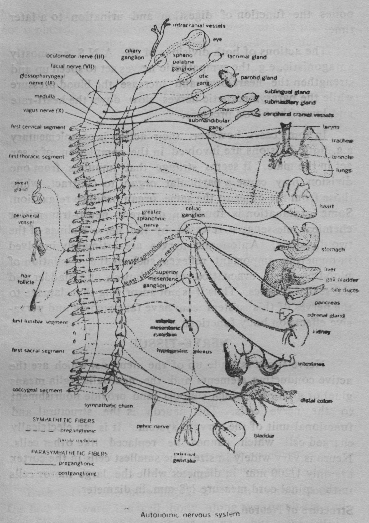
When an emergency arises, the sympathetic division switches the body into high gear. Its function is as follows:
It stimulates the secretion of adrenaline and other 'fight or flight' hormones. It stimulates the liver to convert glycogen into glucose to provide the need for quick energy. It shunts away blood from the digestive organs to provide greater supply to the heart and skeletal muscles. It postpones the function of digestion and urination to a later time.
The actions of both divisions of the A.N.S. are mostly antagonistic, e.g. the sympathetic nerves act to speed up and strengthen the heart-beat and increase the blood pressure while the parasympathetic nerves slow down the heart-rate and lower the blood pressure.
In some cases, however, their action is complementary e.g. both divisions are involved in the completion of a sex act in the male. It seems strange that impulses from one division may cause a particular organ to contract, while those from the other to the same organ produce relaxation. Some explanation is found in different neuro-transmitter chemical messengers released by the nerve-endings of the two divisions. Autonomic nerves are intimately involved in numerous important reflexes such as the regulation of blood pressure, evacuation of the bowels and bladder and the sexual reflexes. Thus it is not surprising that damage to the nerves of this system can have far-reaching and pervasive effects on body functions.
NERVE-TISSUE
Nerve-tissue is made up of the neurons, which are the active conducting elements and the neuroglia (glia means glue) or glial cells which support and provide nourishment to the rerve cells. The neuron is the structural and functional unit of the nervous system. It is an electrically charged cell which cannot be replaced like other cells. Neurons vary widely in size. The smallest cells in the cortex are only 1/200 mm. in diameter while the large motor cells in the spinal cord measure 1/8 mm. in diameter.
Structure of Neuron
Neurons are highly specialized in two key areas of function: excitability and conductivity. A typical neuron consists of a cell body and two types of protoplasmic extensions: axons and dendrites. Some of the neurons are as much as three feet long. Each neuron has only a single axon, but most of them possess numerous dendrites. If the cell body is damaged, the extensions are unable to conduct impulses and the neuron dies. Moreover, the neuron is incapable of dividing itself and when a neuron dies, it is not replaced. All neurons in a body were already present at birth. No new ones are added, but considerable number might be lost.
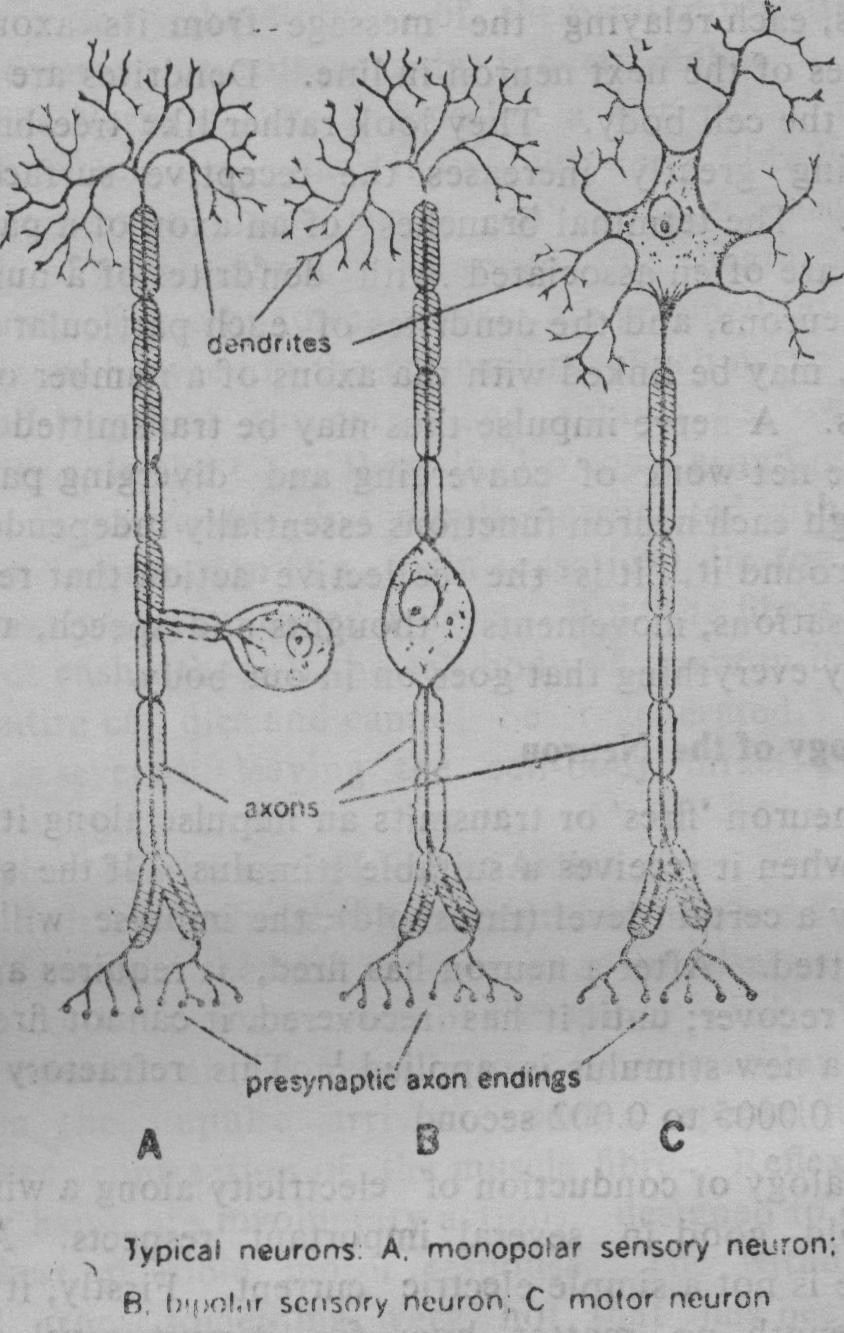
Each neuron conducts impulses in only one direction. The flow is toward the cell body along the dendrites and.away from the cell body along the axons. Thus two separate sets of neurons are necessary for the two-way traffic of messages in the peripheral nervous system. Thus a functional classification of neurons yields two [4] main groups viz., (i) sensory or afferent neurons transmit impulses from the periphery to the C.N.S. and (ii) motor or efferent neurons transmit messages from the C.N.S. to a muscle, a gland or some other tissue and produce an action. In some cases the impulse is transmitted along a chain of motor neurons, each relaying the message from its axon to the dendrites of the next neuron in line. Dendrites are extensions of the cell body. They look rather like tree-branches. Branching greatly increases the receptive surface of the neuron. The terminal branches of an axon of a particular neuron are often associated with dendrites of a number of other neurons, and the dendrites of each particular neuron in turn, may be linked with tha axons of a number of other neurons. A nerve impulse thus may be transmitted over an intricate net-work of converging and diverging pathways. Although each neuron functions essentially independently of those around it, it is the collective action that results in our sensations, movements, thoughts and speech, affecting virtually everything that goes on in our body.
Physiology of the Neuron
A neuron 'fires' or transmits an impulse along its entire length when it receives a suitable stimulus. If the stimulus is below a certain level (threshold), the impulse will not be transmitted. After a neuron has fired, it requires a certain time to recover; until it has recovered, it cannot fire again, even if a new stimulus is applied.[5] This refractory period is from 0.0005 to 0.002 second.
Analogy of conduction of electricity along a wire does not hold good in several important respects. A nerve impulse is not a simple electric current. Firstly, it retains its strength, no matter how far it must travel, for it is continually being renewed as it passes along. Secondly, the speed of nerve transmission is much slower, only about 20 metres per second as against the speed of electric current transmission which is 3,00,000 k.m./sec. (=velocity of light).
The Synapse: The successive neurons in a nerve tract are not directly connected, The terminal branches of an axon are separated from the dendrites or cell body of the next neuron in the sequence by a small gap (200 to 300°A) called the synapse. Nerve impulses are transmitted across the synapse by the diffusion of chemical transmitters. The most common neurotransmitter is acetylcholine.
The Nerve: Like a muscle, a peripheral nerve is a bundle of bundles. Each individual nerve fibre is enclosed in a connective tissue sheath called the endoneurium. A bundle of nerve fibres is wrapped in a sheath called perineurium. These bundles, in turn, are grouped into a larger bundle ensheathed in the epineurium. Each nerve fibre runs the full length of the nerve and retains its own distinct identity. In the CNS, there is also some grouping of nerve fibres, fibres transmitting impulses associated with a specific type of information (e.g. pain-sensation) lie together in a common pathway called a tract. But the fibres of a tract are not ensheathed. If the cell body of a neuron is damaged, the entire cell dies and cannot be regenerated. But if the axon is severed, leaving the cell-body intact, a new axon may be regenerated.
Voluntary Movement and Reflex Action
Willed or voluntary movement begins when the neurons of the cerebral cortex send nerve impulses along nerve fibres to the motor neurons of the spinal cord. From here the impulses are conveyed to neuro-muscular junctions. When the impulse arrives at such a special junction, it induces contraction of the muscle fibre. Reflexes, on the other hand, are involuntary actions, designed to obtain the quickest possible motor response. The withdrawal of a hand from something very hot that has been touched inadvertently is an example of reflex action. It is a protective mechanism of the central nervous system having an incoming sensory and an outgoing motor pathway in common i.e the incoming message is carried directly to the motor-neuron in the spinal cord. The spinal cord and the brain stem are responsible for most of the reflex movements and the use of cerebral cortex is not involved. They are inborn and involuntary actions and they may still be present even after a great deal of brain damage has occurred.
Normally muscular movements are smooth and coordinated because of reflex activity, nervous feedback systems and the mass of information impinging on the spinal motor neurons. If the normal working of the brain and spinal cord is disturbed, movements may become weak, stiff or jerky and actions like walking may become impossible
The cerebellum is important in regulation of movement and in maintaining balance. It works in close connection with the balance organs called labyrinths. They are situated in the inner ears, deep in the sides of the skull one on each side. They pick up information about the position of the head, analyse it, and send messages to the muscles of the body to help preserve balance.
A familiar illustration of the distinction between sensation and perception is 'pain'. The same sensation signal from a toothache, for example, effects each person differently. His suffering is perception. This distinction is utilized as an instrument for developing freedom from the vitiating emotions of like and dislike in the technique of Sharira-Preksha.
The twelve cranial nerves are: I. olfactory, II. optic, III. oculomotor, IV. trochlear, V. trigeminal, VI. abducens, VII. facial, VIII. acoustic, IX. glossopharyngeal, X. vagus, XI. spinal accessory and XII. hypoglossal nerves.
There is a third group of neurons referred to as inter-neurons. They are restricted to the C.N.S. and relay impulses to various functional centres in the brain or spinal cord.