 Jethalal S. Zaveri
Energy is essential to maintain such vital functions and processes as breathing, blood circulation and brain function. It is derived from foodstuffs which can be divided into several classes. Most of the food for human consumption is derived from vegetables and animals. Since they have already built up the complex molecules of proteins, carbohydrates and lipids (fats). Food materials cannot be utilised by the tissues until they have been broken down to smaller components and turned into a soluble form in order to reach the cells through the process of digestion. The digested materials can then be absorbed and sent to the tissues for production of energy.
Digestive Processes
Two types of actions take place in various digestive organs: -
- Mechanical actions
- Chemical actions
Mechanical Actions. Grinding, mixing and churning he food play an important part in digestion. Mere mechanical actions are, however, not enough. They move the food materials along, moisten and liquefy them and pulverize them and thus prepare the way for chemical actions of the digestive enzymes in various organs.
Chemical Actions. They convert the large and complicated molecules of the foodstuff to smaller simpler units so that they can be extracted from the digestive organs by the blood vessels, sent into the blood stream and can pass through cell membranes for assimilation by the tissues. Water, mineral oils and vitamins do not undergo chemical action in order to be absorbed.
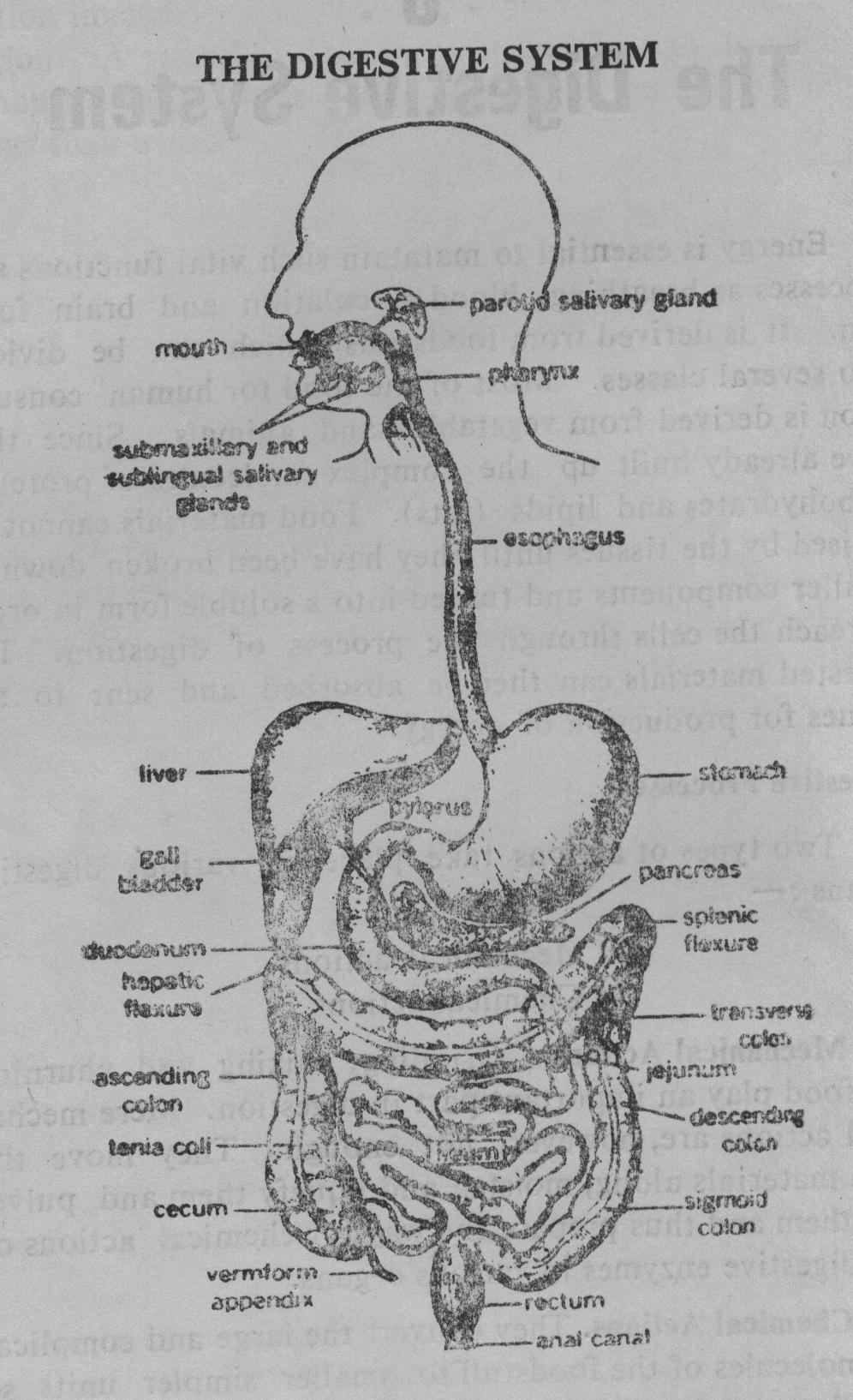
Organs of the Digestive System
The digestive system is composed of the alimentary canal and accessory organs which contribute their secretions to the tract. The digestion of food begins in the mouth and ends in the bowel. The passage (tract) from the mouth to the rectum (the end portion of the bowel) is called gastro-intestinal tract which is like a tube, about 9 metres in length, elaborately looped and coiled within the body. Various processing stations are located along the way to prepare and process the food and absorb useful materials The accessory organs of the digestive system include salivary glands, the pancreas, the liver and gall bladder.
1.Mouth and Salivary Glands
The first station on the route of the alimentary canal is mouth. Food spends possibly the shortest time of the whole process in the mouth. Nevertheless, it is one of the most important parts of the digestive process, since it is here that the food undergoes its first stage of conversion into a substance which can eventually be more readily absorbed by the cells of the body. Solid food token into the mouth is first broken up into small pieces and then chewed and ground to pulp by the teeth. Grinding the food up is essential to increase the surface area of the food. Saliva enters the mouth through ducts from three pairs of salivary glands which are accessory organs of the digestive system. Saliva contains the enzymes, one of which is ptyalin which starts off the chemical breakdown of starchy foods into sugar. The tongue pushes the food between the teeth, shapes masticated food into a convenient bolus and shoves it downwards to the aesophagus.
2. Aesophagus
The ground food-mass, lubricated and softened by saliva, passes backwards on the epiglottis into the aesophagus which is a muscular tube about 2.5 cms. in dia and 25 cms. long, leading down through the chest and diaphragm to the stomach. It is a flexible tube and if the miss of food is larger than its diameter, it can expand to accommodate it. Swallowing the moist ball of food is a chain of complicated movements, involving operation of reflex mechanisms to ensure that the food will go where it is meant to. First the soft palate rises blocking off the nose and prevent food from entering the nasal cavity. Both the aesophagus and trachea open into the mouth. The valve called epiglottis guards the trachea during the swallowing process, preventing the food from entering the trachea. However, sometimes, inadvertently, this automatic guarding mechanism may fail and the portion of the food may enter the trachea, which is usually thrown out by a typical fit of coughing. If, however, a piece of food gets lodged into the trachea, it creates hazard to life and must, therefore, be removed quickly. Once the food enters the aesophagus, it is propelled down to the stomach by alternate waves of contraction and relaxation of the muscular tube.
3. The Stomach
The food has now reached the second processing station of the tract. The muscular collapsible bag called stomach is tucked up in the abdomen at the lower rib-line under the diaphragm and liver, and resembles a deflated balloon when empty. Its size and shape vary depending on how full it is. Its average size is about 22 cms. in length. When full, it slants across the body, big at the top and small at the bottom. Its capacity is two to three pints. It retains food for several hours, during which time partial digestion of protein takes place. Like the mouth, the stomach performs its function by both mechanical and chemical actions. The muscular contractions of the stomach act, like a churn and mix food thoroughly with the digestive juices. They toss, turn and mix the stomach contents, gradually macerating the food materials, mixing them with the acid gastric juice and converting them to a semi-fluid mixture. The lining of the stomach contains 35 million gastric glands which secrete four to five pints (2 to 3 litres) of gastric juice per day. Gastric juice contains mucin, hydrochloric acid and enzymes pepsin (the main component) and rennin.[1] Hydrochloric acid is very important in digestion as it activates enzymes (biological catalysts), helps in the digestion of protein and destruction of bacteria. It continues to be produced even when the stomach is empty. Rennin acts on the milk and separates its solid portion. Thereafter, in the presence of the acid, pepsin breaks down the proteins to polypeptides which would be finally digested in the intestine Some form of sugar is absorbed by the blood-vessels from the stomach itself. Mucin lines the stomach and protects it from the acid and prevents it from digesting itself. Gradually the thick gruellike mixture of food and digestive juices called chyme is pushed by vigorous peristaltic waves of the stomach towards the pyloric valve which opens into duodenum, the first part of the small intestine. The function of the pyloric valve is to restrict the admittance of the highly acidic mass into the duodenum—no more than can be instantly neutralized by alkali arriving from pancreas and liver. Peristalsis is the name given to the slow automatic movement of the stomach and also along the whole length of the gastrointestinal tract, propelling the content onwards- After the stomach has been empty for a long time, intense rhythmic hunger-contractions may sweep over its body.
4. Small Intestine
The stomach extends from the aesophagus to the duodenum, the first portion of the small intestine. The lower end of the stomach which becomes considerably narrower is connected to the small intestine through pyloric valve. The small intestine which is a coiled twisted mass in the centre of the abdomen is divided into three parts: (i) duodenum (adjacent to the stomach), (ii) jejunum and (iii) ileum, totalling more than 7 metres. It is the third processing station of the digestive track. Like the stomach, it mixes and moves the food. It is here that the main work of digestion and about 90% of the absorption of the food constituents into the blood-stream takes place. A variety of enzymes and other digestive substances secreted not only by the small intestine but also by the liver and pancreas, act here and complete the breakdown of proteins, carbohydrates and fats into simple constituents. Here too, the major work of absorption takes place and nutrients are absorbed through the intestinal wall into the blood-circulation. Normally, 3 to 10 hours are required for chyme to pass from the pylorus to the end of the ileum.
(a) Duodenum. The first part of the small intestine is about 16 to 17 cms. long. A 'C-shaped' tube, the duodenum begins at the pylorus, passes behind the liver in front of the right kidney and across the aorta. It encircles the head of the pancreas.
The structure of this organ is similar to that of the stomach. The only difference is that its inner lining is very much wrinkled, resulting in greater surface area in a smaller space. This is an important organ in the digestive system. The pancreatic juice and the bile enter the tract about 7 cms. from the pylorus and commence their digestive action on food. Also, here—but not elsewhere—are glands, called the Brunner's glands, beneath the mucous membrane. Small quantities of thick gruel-like mixture of semi-digested food and gastric juices are squirted into the duodenum through the gate-keeper like valve. Since this is highly acidic, too much at a time would damage the lining of the duodenum, which is notorious as a site of ulcer, since it bears the brunt of jets of acidic chyme that periodically squirt into it from the stomach. To neutralize most of the acid, alkaline digestive juices from the pancreas and gall bladder pour into the duodenum via the pancreatic duct with the characteristic split-second timing and meet the chyme. These juices contain three main enzymes which separate the proteins, fats and carbohydrates into basic building blocks.
(b) Jejunum and Ileum. There is no clear demarcation between jejunum and ileum, although their membranes differ somewhat in structure. The jejunum constitutes about two-fifths of the small intestine and the somewhat longer ileum, the rest. The ileum ends in a right angled T-junction with the large intestine. The greater part of food-digestion and absorption takes place in the jejunum and the ileum. The slightly acidic liquid which enters the jejunum leaves it as an alkaline one. During its passage, virtually all t'e nutrient materials are extracted.
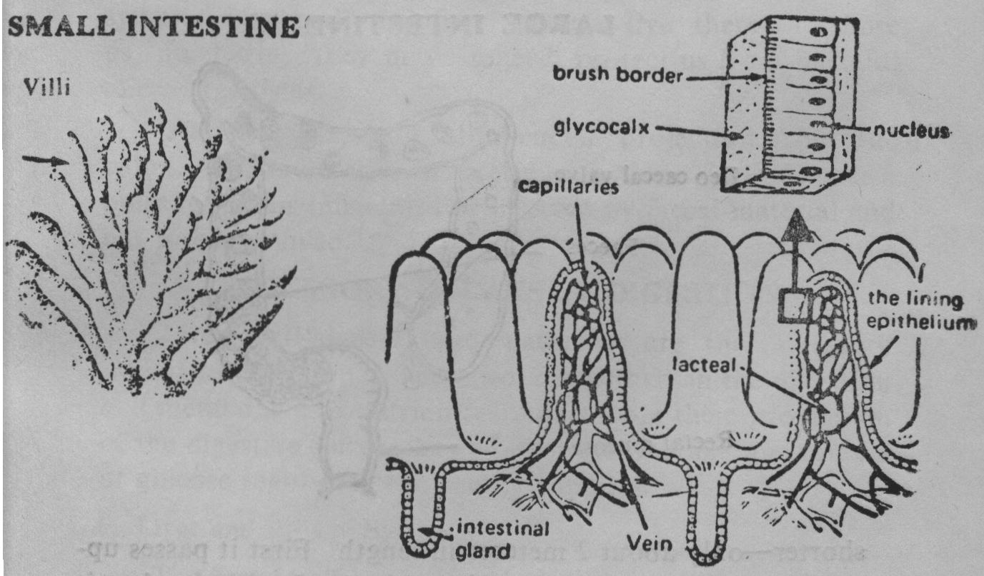
The Absorption Process
The structure of the small intestine is specialized so that the absorption of nutrients can proceed most efficiently. The absorption of valuable nutrients results from efficient enzyme action and the way with which the intestinal lining absorbs them from the cavity of the intestine. Perhaps the most important components of the intestine are millions of villi—tiny finger-like projections on its walls. These are the working structures of the small intestine in its function of absorption of digested nutrients. Each villus contains a network of capillaries, and a lymph vessel. (See, figure). Nutrients pass through into the small blood-vessels which run into larger ones and eventually into the hepatic vein which leads to the liver, where breakdown continues, before final delivery to the other cells of the body. Once absorbed by the body, the broken-down substances provide the raw materials for the building of longer and more complicated molecules which are better suited to the needs of the body.
The end products of digestion which are absorbed include glucose (from carbohydrates), amino-acids (from proteins) and a milky fatty emulsion. They are put into circulation—glucose and amino-acids via the blood-stream and fatty emulsion via the lymphatic system. Over and above these three main nutrients, the following are also extracted and made available for bodily functions:
- Salts and Minerals
Calcium, Potassium, Sodium, Iron, Phosphorus, Iodine.
-
Vitamins
- Soluble in fats
- Soluble in water
- Water
Out of these, absorption of vitamins generally takes place in small intestine, while salts and water are absorbed in large intestine. What remains of the food after assimilation then passes into the large intestine, where the final stage of the digestive process takes place.
5. Large Intestine or Colon

Next to the small intestine comes the large intestine or bowel. It is much wider - 6 to 8 cms. in diameter—but shorter—only about 2 metres in length. First it passes upwards, and is called 'ascending colon'. It bends when it reaches the bottom of the liver and remains horizontal upto the spleen. This is known as transverse colon. It, then, bends downwards and is called 'descending colon'. Its last part is in the pelvic cavity and is called the rectum, which is about 25 cms. long, and is rich in muscular tissues. Finally it ends at the external opening, the anus. The obvious differences from the small intestine are: larger diameter, a puckard rather than tube-like outside, and the longitudinal muscle being arranged in bands. The lining secretes mucus, but no digestive enzymes.
This is the fourth and last processing station of the tract. By the time it is reached, almost all the useful nutrients have already been digested and absorbed. What remain are indigestible stuff together with salts, bile-pigments, and large valuable quantities of water (the digestive juices are about 95% water). The function of the large bowel is extraction of salt and water (that can be usefully recycled) and the formation of the faeces from the indigestible food-stuffs like cellulose, bowel secretions and bacteria. The lower parts contain many organisms which manufacture a variety of useful substances including vitamin-K and several vitamins of B group as well as the smelly compounds responsible for the odour of faeces. Not all the inhabitants of our colon are hostile parasites. Millions of symbiotic (mutually helpful) bacteria live there and cause us no harm; they may indeed protect us from harmful micro-organisms.
The appendix is a blind-ended projection from the colon. It has no known useful function, but becomes a nuisance when inflamed or infected by faecal material and has to be removed.
ACCESSORY ORGANS OF DIGESTION
Liver, gall-bladder and pancreas are the accessory organs of digestion. They work together in the digestion and metabolism of nutrients. Apart from their production of the digestive juices, they control the storage and release of glucose regulating the energy supplies.
6. Liver and Biliary System
The liver itself is an amazing chemical factory within the body, generally underrated and abused. Even when badly damaged, its cells have enormous powers of regeneration.

The liver is the most important and an extraordinary organ which contributes to the process of digestion outside the gastro-intestinal tract. It is not only the largest but the most versatile single organ in the body. It lies in the upper part of the abdomen on the right side beneath (and loosely attached to) the diaphragm. It is reddish brown in colour and weighs about 1.5 to 2 kg. A remarkable feature of the liver is that it receives a double supply of blood: (a) fresh arterial blood from the hepatic artery arising from the aorta, and (b) from the portal vein carrying the finished products of digestion from the intestine. Both these large bloodvessels branch repeatedly into a network of thousands of tiny capillaries making the liver a highly vascular organ.
Blood from both the blood-vessels mixes in the liver through its kupffer cells. Blood from liver carrying the nutrients reaches the vena cava through the hepatic vein. Thus nutrients processed by the liver reach the blood-circulation through vena cava.
Bile-ducts and Gall-bladder
Production of bile is only ore of the many functions of the liver cells. Bile contains 86% water, bile-salts, bile-pigments and cholesterol etc. It aids in the emulsification and digestion of fats.
The ducts carrying the bile secreted by the liver to the gall-bladder are called bile-ducts. Bile is a thick, dark-green, alkaline digestive juice Attached to the lower surface of the liver is a small blind pear-shaped pouch called the gall-bladder which receives, stores and concentrates bile. The gall-bladder is far too small to hold the 1000 cc of bile produced daily by the liver, so it has the ability to concentrate the bile up to 20 times. When required, bile from the gall-bladder passes into duodenum together with the pancreatic juices through a common duct. Bile-salts are responsible for breaking globules of fats into tiny droplets.
The bile-salts are not lost alter fulfilling their role in the digestive process, but are carried back to the liver to be resecreted into the bile. This illustrates the high efficiency of the system which recycles the very small amount 13 to 4 gms) of bile-salts present in the adult.
Functions of Liver
Liver is the largest chemical factory in the body, and has at least five hundred known functions. It is an exocrine gland.[2]
All the absorbed nutrients from digestion pass through this biochemical factory.
The cells of the liver contain a variety of enzymes for many chemical processes, and they are also the vital stores of essential material. The metabolism of each of the three groups of food-material, viz., carbohydrates, fats and proteins takes place in the liver.
If the body-cells require immediate energy, the liver releases some of the glucose back into the blood-stream for delivery to the cells. The remaining glucose is converted into glycogen—a larger molecule—which can be stored in the liver and some muscle cells. When all the glycogen storage areas are filled up, the remaining glucose is converted into fat and stored. The liver has the proper enzymes which are necessary to carry out the conversions of carbohydrates, proteins and fats into one another.
Another function of the liver is to store important vitamins including A, D and B12 and iron.
The liver neutralizes the injurious effects of the toxics such as poisonous drugs and liquor. Besides, whenever a toxic substance reaches the liver from the intestines etc, it is processed in the liver, and rendered excretable through bile or urine. However, the poison of excessive drinking or hypnotic drugs could destroy the liver cells.
Yet another function of the liver is to produce urea-When amino-acids absorbed by the blood-vessels in the villi (in the intestine) reaches the liver-cells, it is de-aminized, i.e. nitrogen is separated and transformed into urea which is excreted through urine by the kidneys.
Over and above the above-mentioned functions connected with the digestive system, the liver has to perform some important functions pertaining to the general components of blood. For example, (i) production of new red blood-cells during the foetal life, and (ii) assistance in maturing them later on and withdrawing them from the circulation on their becoming worn-out, (iii) breaking-down of haemoglobin from the worn-out red blood-cells and converting it into an iron containing pigment, bile-pigments—bilirubin and biliverdin (while most of the iron is reutilized, bilirubin etc. is excreted).
Lastly, it also assists in keeping the body-temperature constant.
7. Pancreas and the Islets of Langerhans
Pancreas is the most important producer of digestive juices in the whole digestive system. It is a versatile organ, the second largest gland in the body (the largest is the liver), and functions both as a digestive organ (exocrine gland), and as an endocrine gland. In fact, it is two unrelated organs into one.
The pancreas is an oblong, rather flattened, boneless, fatless and fleshy gland about 15 cms. long, grey-pink in colour, and weighs about 85 gms. It consists of a head, a body and a tail; its head rests in the curve of the duodenum and its tail touches the spleen. The pancreas is connected to the duodenum through the pancreatic duct which extends throughout its length.
Scattered throughout the pancreas, between the glands that pour juice into the pancreatic duct, are many pin-head-sized clusters of special cells. These are the islets of Langerhans.
Exocrine Function
The exocrine portion of the pancreas produce 1000 to 1200 ml (2 pints) of alkaline fluid daily. This pancreatic juice contains several digestive enzymes.
Highly acidic gruel which leaves the stomach can spell disaster in the digestive tract by eating away the delicate lining of the small intestine (duodenum). To neutralize it, the pancreas (in association with the gall-bladder which sends the bile to duodenum) must produce enough alkaline juice. It waits on call, ready to supply a powerful arsenal of digestive enzymes as soon as they are needed. It does not begin to pour its products into the duodenum until food reaches there. Commencing its action through nervous control, the pancreas is prodded into full action by the chemical message of the hormone secretion produced by the duodenum as a measure of self-protection. These hormones stimulate the pancreas to produce juice rich in sodium bicarbonate which neutralizes the acid of the chyme.
Pancreatic juice has five main enzymes. Three of these complete the digestion of proteins begun in the stomach. The others are (i) amylase (which digests carbohydrates), and (ii) lipase, the only fat-digesting enzyme in the body, which works on the tiny fat droplets prepared by the bile.
Endocrine Function
The endocrine portion (the Islets of Langerhans) of the pancreas contain two main types of secreting cells, alpha and beta cells which secrete the hormones—glucagon and insulin. While many people may have a rough idea of what insulin does, only a few would have ever heard of glucagon. Both glucagon and insulin are concerned with the regulation of the body's carbohydrate metabolism, but their effects are opposite. (Yet the two hormones do not antagonize or block one another; they work independently). When the blood-sugar-level rises after a meal, for example, the secretion of insulin is stimulated, and it causes the blood-sugar-level to fall. When the blood-sugar-level falls below the normal value, glucagon is secreted which raises the glucose-level of the blood. Thus, insulin and glucagon together keep the blood-sugar-level within a relatively narrow range.
Insulin acts in several ways to lower the blood sugar-level. It facilitates the transport of glucose through cell-membranes, since the rate at which a cell utilizes glucose is determined to a large extent by the rate at which it enters the cell. Insulin thus speeds up the rate of glucose metabolism. It also acts on the cellular enzymes that catalyze the conversion of glucose to glycogen, and thus helps to take glucose out of circulation and store it away.
Insulin also stimulates synthesis of fatty acids from glucose and inhibits the conversion of amino and fatty acids to glucose in the liver.
Failure to produce insulin in adequate quantities results in diabetes mellitus, i.e. increase of blood-sugar which appear in the urine. In the absence of insulin, cells would try to burn fat and/or protein which would have to be drained from muscle tissues, while unburned sugar would pass out of the body in sweetish urine taking with it a lot of water and useful salts.
Consumption of sweets in excess amount requires increased insulin production.
Thus, the so-called accessory organs perform a phenomenal multiplicity of biochemical functions unequalled by any other organs.
Metabolism
Metabolism is the name given to the biochemical processes consisting of the breakdown of the basic food-materials with the release of energy and their re-arrangement into complex substances which build up the living tissues. It involves the digestion of food in the stomach and intestines, the absorption and storage of digested materials, their incorporation into the tissues of the body and finally their release and breakdown to water and carbon dioxide with the liberation of energy [3].
lnfants and children whose main diet is milk have a special need for the abilities of rennin. It is not a very important component in the dault.
Glands producing secretions, which drain out through ducts and have an effect only near the area where they are released, are called exocrine (exo=outside), in contrast to endocrine (endo=inside) or ductless glands manufacturing hormones which pass directly into the blood-stream, circulate all over the body and at places far from where they are secreted. For further details, see later section.