 Prof. J.P.N. Mishra
Skeletal muscle (Fig 1-20A) constitutes approximate 40% of the body's weight and is responsible for locomotion, facial expression, posture, respiratory movements, and many other body movements. The nervous system voluntarily, or consciously, controls the functions of the skeletal muscles.
Smooth muscle (Fig 1-20B) most widely distributed in the body having greatest variety of functions, are found in the walls of hollow organs and tubes, the interior of the eye, the walls of blood vessels, and other areas. Smooth muscle performs a variety of functions, including propelling urine through the urinary tract, mixing food in the stomach and intestine, dilating and constricting the pupils, and regulating the flow of blood through blood vessels.
Cardiac muscles (Fig 1-20C) are found only in the heart, and their contractions provide the major force for heat beat. Unlike skeletal muscle, cardiac muscle and many smooth muscles are autohythmic. They contract spontaneously at somewhat regular intervals. Unlike skeletal muscle, smooth muscle and cardiac muscle are not consciously controlled by the nervous system. Rather, they are controlled involuntarily, or unconsciously by the autonomic nervous system and the endocrine system.
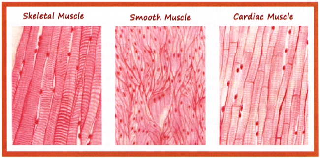
Fig 1-20. Muscle tissues. (A) Skeletal. (B) Smooth. (C) Cardiac.
Membrane Potentials: Plasma membranes are remain polarized means there is a voltage difference, or electrical charge difference, across the membrane before action potentials can be generated. This charge difference is called the resting membrane potential. The negative charge at the internal surface of the plasma membrane compared to its outer surface results mainly from the concentration differences of ions and charge molecules across the plasma membrane and to its permeability characteristics. The concentration of K+ inside the cell is much higher than its concentration outside the cell. The plasma membrane is relatively permeable to K+ and much less permeable to negatively charge molecules found inside the cell. Consequently, positively charge K+ tends to diffuse out of the cell leaving the negatively charged molecules behind. The membrane becomes polarized when the tendency for K+ to diffuse out of the cell is resisted by the negative charges of the molecules inside the cell.
Ion Channels: Once the resting membrane potential is established, action potentials can be produced. An action potential is a reversal of the resting membrane potential such that the inside of the plasma membrane becomes positively charged compared to the outside. The permeability characteristics of the plasma membrane change as a result of the opening of certain ion channels, when a cell is stimulated. The diffusion of ions through these channels changes the charge across the plasma membrane and produces an action potential.
Action Potentials: An action potential takes j approximately to a few milliseconds to occur, and it has two phases called depolarization and repolarization. Stimulation of a cell can cause depolarization of its plasma membrane, which is graphed. Depolarization occurs when the inside of the plasma membrane becomes less negative, which is indicated by movement of the curve upward toward zero. The depolarization phase of an action potential is triggered if the depolarization changes the membrane potential to a value called threshold. The charge difference across the plasma membrane reverses when the membrane potential becomes a positive value. Repolarization is the return of the membrane potential to its resting value (Fig. 1-21).

Fig 1-21. Electrical charges and ion concentrations at the sarcolemma.
Neuromuscular Junction: Motor neurons with the help of their axons carry action potentials at a high velocity from the brainstem and spinal cord to skeletal muscle fibres. The axons branch repeatedly, and each branch projects toward one muscle fibre to innervate it. Thus, each muscle fibre receives a branch of an axon, and each axon innervates more than one muscle fibre (Seelay...). Near the muscle fibre it innervates, each axon branch forms a cluster of enlarged axon terminals that rests in an invagination of the sarcolemma to form a synapse, or neuromuscular junction, which consists of the axon terminals and the area of the muscles fibre, sarcolemma they innervate. Each axon terminal is the presynaptic terminal. The space between the axon terminal and the muscle fibre is the synaptic cleft, and the muscle plasma membrane in the area of the junction is the postsynaptic membrane, or motor end- plate.
Each presynaptic terminal contains numerous mitochondria and many small, spherical sacs approximately 45 pm in diameter called synaptic vesicles. The vesicles contain acetylcholine, an organic molecule composed of acetic acid and choline, which functions as a neurotransmitter. A neurotransmitter is a substance released from a presynaptic membrane that diffuses across the synaptic cleft and stimulates (or inhibits) the production of an action potential in the postsynaptic membrane (Fig. 1-22).
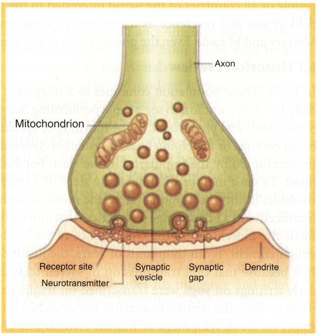
Fig 1-22, Structure of the neuromuscular junction, showing an axon terminal adjacent to the sarcolemma of a muscle fibre.
Cross-Bridge Movement: A cycle of events resulting in contraction proceeds very rapidly when the heads of the myosin molecules bind to actin. The heads of myosin molecules move at their hinged region, forcing the actin myofilament, to which the heads of the myosin molecules are attached, to slide over the surface of the myosin myofilament. After movement, each myosin head releases from the actin and returns to its original position. It can then form another cross-bridge at a different site on the actin myofilament, followed by movement, release of the cross-bridge, and return to its original position. During a single contraction, each myosin molecule undergoes the cycle of cross-bridge formation, movement, release, and return to its original position many times.
Muscle Relaxation: Relaxation occurs as a result of the active transport of Ca2+ back into the sarcoplasmic reticulum. As the Ca2+ concentration decreases in the sarcoplasm, the ions diffuse away from the troponin molecules. The troponin-tropomyosin complex then re-establishes its position, which blocks the active sites on the actin molecules. As a consequence, cross-bridges cannot re-form once they have been released, and relaxation occurs. Energy is needed to make muscles contract, but it is also needed to make muscles relax. The active transport of Ca2+ into the sarcoplasmic reticulum requires ATP. The active transport processes that maintain the normal concentrations of Na+ and K+ across the sarcolemma also require ATP. The amount of ATP required for cross-bridge formation during contraction is much greater than the other energy requirements in a skeletal muscle.