 Prof. J.P.N. Mishra
The central nervous system (CNS) consists of the brain and the spinal cord and is protected by bone: The brain occupies the cranial cavity, and the spinal cord occupies the vertebral canal.
Brain: The human brain is divided into Cerebrum, Diencephalon, Brain Stem and Cerebellum. The brain has four cavities, called ventricles: two lateral ventricles, the third ventricle;-and the fourth ventricle. Interventricular foramina connect the lateral ventricles with the third ventricle. The third and fourth ventricles are connected by a small canal called the cerebral aqueduct (aqueduct of sylvius).
Brain Stem: The medulla oblongata, pons, and midbrain are the subdivisions of brain stem. The medulla oblongata lies between the spinal cord and the pons and is anterior to the cerebellum. It contains a number of vital centers for regulating heartbeat, breathing, and blood pressure. It also contains the reflex centers for vomiting, coughing, sneezing, hiccoughing, and swallowing. The medulla contains tracts that ascend or descend between the spinal cord and the brain's higher centers.
The pons is a "bridge;" between the cerebellum and the rest of the CNS and it functions with the medulla to regulate breathing rate and has reflex centers concerned with head movements in response to visual and auditory stimuli.
Cerebellum: The cerebellum (Fig 1 -16) lies below the posterior portion of the cerebrum, is separated from the brain stem by the fourth ventricle. The surface of the cerebellum is gray matter, and the interior is largely white matter. The cerebellum functions are muscle coordination, integrating impulses received from higher centers to ensure that all of the skeletal muscles work together to produce smooth and graceful motion. The cerebellum is also responsible for maintaining normal muscle tone and transmitting impulses that maintain posture. It receives information about body position from the inner ear and then sends impulses to the muscles, whose contraction maintains or restores balance.
Functions of the cerebellum
The cerebellum coordinates three important functions, which all are related with the control of skeletal muscles:
- It acts with the cerebral cortex to produce skilled movements by coordinating the activities of groups of muscles.
- It helps controlling the body posture. It functions below the level of consciousness to make movements smooth instead of jerky, steady instead of trembling, and efficient and co-ordinated instead of ineffective, awkward, and uncoordinated.
- It controls skeletal muscles to maintain balance.
Organised control of muscle action, is closely associated with cerebral motor activity. Normal muscle action involves groups of muscles, the various members of which function together as a unit. In any given action, for example, the prime mover contracts and the antagonist relaxes but then contracts weakly at the proper moment to act as a brake, checking the action of the prime mover. Also, the synergists contract to assist the prime mover, and the fixation muscles of the neighbouring joint contract. Through such harmonious, coordinated group action, normal movements are smooth, steady, and precise as to force, rate, and extent.
Net outcome of such movements results from cerebellar activity. Impulses from the cerebrum starts the action, but those from the cerebellum synergize or coordinate the contractions and relaxations of the various muscles once they have begun. Impulses from the motor control areas of the cerebrum travel down the corticospinal tract and, through peripheral nerves, to skeletal muscle tissue. At the same time, the impulses go to the cerebellum. The cerebellum compares the intended movement to the actual movement. Impulses then travel from the cerebellum to both the cerebrum and the muscle tissue to adjust or coordinate the movements to produce the intended action. In view of various physiologists it is main function of the cerebellum. One part of the cerebellum is also thought to be concerned with both exciting and inhibiting postural reflexes. In addition, part of the cerebellum presumably discharges impulses important to the maintenance of balance. Sensory impulses from equilibrium receptors in the ear reach the cerebellum. Here, connections are made with the proper motor fibres for contraction of the necessary muscles for stabilizing the body.
The midbrain encloses the cerebral aqueduct. Aside from acting as a relay station for tracts passing between the cerebrum and the spinal cord or cerebellum, the midbrain has reflex centers for visual, auditory, and tactile responses.
Diencephalon: The hypothalamus and thalamus constitute diencephalon, where the third ventricle is located. The hypothalamus, which forms the floor of the third ventricle. It maintains homeostasis, or the constancy of the internal environment, and contains centers for regulating hunger, sleep, thirst, body temperature, water balance, and blood pressure. The hypothalamus also controls the pituitary gland and thereby serves as a link between the nervous and endocrine systems.
The thalamus, in the lateral walls of the third ventricle, is the last portion of the brain for sensory input before the cerebrum. It functions as a central relay station for sensory impulses traveling upward from other parts of the body and brain to the cerebrum. It receives all sensory impulses (except those associated with the sense of smell) and channels them to appropriate regions of the cortex for interpretation (Saladin, 2004).
Thalamus: The thalamus is a dumbbell-shape structure made up of gray matter. Extending through the third ventricle, and thus joining the two lateral masses of the thalamus, is the intermediate mass. Two important groups of nuclei comprising the thalamus are the geniculate bodies, located in the posterior region of each lateral mass. The geniculate bodies play a role in processing auditory and visual input (fig. 1-16).
Large numbers of axons conduct impulses into the thalamus from the spinal cord, brainstem, cerebellum, cerebral nuclei, and various parts of the cerebrum. These axons terminate in thalamic nuclei, where they synapse with neurons whose axons conduct impulses out of the thalamus to virtually all areas of the cerebral cortex. Thus the thalamus serves as the major relay station for sensory impulses on their way to the cerebral cortex.
Important functions of the Thalamus are
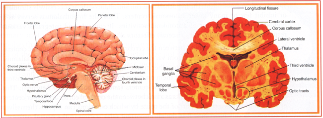
Fig 1-16.(A)Midsagittal section of the brain as seen from the left side. This medial plane shows internal anatomy as well as the lobes of the cerebrum. (B)Frontal section of the brain in anterior view.
- The impulses received from appropriate receptors, on reaching the thalamus, produce conscious recognition of the crude, less critical sensations of pain, temperature, and touch, which are regulated by thalamus.
- Neurons having their dendrites and cell bodies in certain nuclei of the thalamus relay all kinds of sensory impulses, to the cerebrum.
- It plays a part in the mechanism responsible for emotions by associating sensory impulses with feelings of pleasantness and unpleasantness.
- It also plays a part in the arousal or alerting mechanism
- Further it also plays a part in mechanisms that produce complex reflex movements
Hypothalamus: The hypothalamus is consists of several structures that lie beneath the thalamus and form the floor of the third ventricle and the lower part of its lateral walls (Fig 1-16). Structures constituting the hypothalamus are the supraoptic nuclei, the paraventricular nuclei, and the mammillary bodies. The supraoptic nuclei consist of gray matter located just above and on either side of the optic chiasma. The paraventricular nuclei of the hypothalamus are so named because of their location close to the wall of the third ventricle. From the midportion of the hypothalamus, it give rise to the infundibulum, the stalk leading to the posterior lobe of the pituitary gland (neurohypophysis). The posterior part of the hypothalamus consists mainly of the mammillary bodies, which are involved with the olfactory sense (smell).
The hypothalamus is a small component of brain having very specialized characteristics. Although it only weighs approx. 7g (1/4 ox), yet it performs many significant functions for both survival and the enjoyment of life. For example, it functions as a link between the psyche (mind) and the soma (body). It also works as link between the nervous system to the endocrine system. Certain areas of the hypothalamus function as pleasure centers or reward centers for the primary drives such as eating, drinking, and sex. The important functions of hypothalamus maybe summarized as follows:
- It works as a higher autonomic center which means that axon of neurons whose dendrites and cell bodies are located lie in nuclei of the hypothalamus extend in tracts from the hypothalamus to both parasympathetic and sympathetic centers in the brainstem and spinal cord. Impulses from the hypothalamus can simultaneously or successively stimulate or inhibit few or many lower autonomic centers. Thus, the hypothalamus serves as a regulator and coordinator of autonomic activities. It controls and integrates various responses made by autonomic (visceral) effectors all over the body.
- It acts as the major relay station between the cerebral cortex and lower autonomic centers. Neural tracts conduct impulses from various centers in the cortex to the hypothalamus. These impulses then are relayed to other tracts that hypothalamus, these impulses are relayed to other tracts that conduct them on down to autonomic centers in the brainstem and cord and also to spinal cord somatic centers (anterior horn motor neurons). This means that the hypothalamus works as a link between the cerebral cortex and lower centers of body. It provides a crucial part of the route by which emotions can express themselves in changed bodily functions. It is the all-important relay station in the neural pathways that makes possible the mind's influence over the body-sometimes, unfortunately, even to the profound degree of producing "psychosomatic disease." The positive benefits of this mind-body link are the dramatic influences our conscious mind can have in healing the body of various illnesses.
- Few hypothalamic neurons located in supraoptic and paraventricular nuclei synthesize the hormones released by the posterior pituitary gland (neurohypophysis), who play an indirect but important role in maintaining water electrode balance.
- Certain neurons of hypothalamus secrete chemicals releasing hormones, into blood, which circulate to the anterior pituitary gland. Those hormones control the release of certain anterior pituitary hormones-specifically growth hormone and hormones that control hormone secretion by sex, thyroid gland, and the adrenal cortex.
- It also plays a key role in maintaining the sleep-wakefulness state. Presumably it functions as part of an arousal or alerting mechanism.
- It also plays as a crucial role in the mechanism for regulating appetite and therefore the amount of food intake. Experimental and clinical findings indicate the presence of an "appetite center" in the lateral part of the hypothalamus and a "satiety center" in the lateral part of the hypothalamus and a "satiety center" located medially.
- It also acts in prominence for maintaining normal body temperature. Few hypothalamus neurons whose fibres connect with autonomic centers for vasoconstriction, dilation, and sweating and with somatic centers for shivering constitute heat-regulating centers. Marked elevation of body temperature frequently characterizes injuries or other abnormalities of the hypothalamus.
Cerebrum: The cerebral cortex is gray and contains cell bodies and short fibres. The cortex has convolutions known as gyri, which are separated by shallow grooves called sulci and deep grooves called fissures. The cerebrum is almost divided by a deep, longitudinal fissure. At the base of this fissure lies the corpus callosum, a bridge of myelinated fibres that joins the two hemispheres. Each cerebral hemisphere has four lobes: frontal, parietal, temporal, and occipital (Fig 1-17), which are named for the bones that cover them.
Association areas are believed to contain areas for intelligence, artistic and creative ability, and learning. The primary sensory area receives nerve impulses from the sense organs and produces what are termed sensations. The particular sensation produced is related to the area of the brain that is stimulated, since the nerve impulse itself always has the same nature. The primary motor area of the cerebrum initiates nerve impulses that control muscle fibres. The primary sensory area of the cerebral cortex lies in the parietal lobe just posterior to the central sulcus; the primary motor area lies just anterior to the central sulcus.
The motor area for speech, called Broca's area, is located near the base of the primary motor area. Broca's area is usually only found in the left cerebral hemisphere. Damage to this area can interfere with a person's ability to understand words (written or spoken) and to communicate with others. This explains why a stroke is often accompanied by speech problems. Strokes occur when the blood supply to the brain is temporarily halted due to a burst blood vessel or to a blood clot in an artery. Electroencephalograms (EEGs), which record the brain's electrical activity, are examined in the Medical Focus reading on this page.

Fig 1-17. Left cerebral hemisphere showing some of the functional areas.