 Prof. J.P.N. Mishra
Structure: The spinal cord originate from the base of the brain and extends through the foramen magnum of the skull into the vertebral canal and terminates between the first and second lumbar vertebrae. A cross section of the spinal cord reveals the central canal and distinct white and gray areas (Fig. 1-18). The gray matter is centrally located and shaped like the letter H. It is gray because it contains cell bodies and short, unmyelinated fibres. The two posterior projections of the H are the dorsal (posterior) horns, and the two anterior projections of the H are the ventral (anterior) horns. Sensory neurons enter the spinal cord through the dorsal roots; motor neurons exit the spinal cord through the ventral roots. The dorsal and ventral roots join to form the spinal nerves. The white matter fills in the areas around the gray matter. The white matter contains bundles of myelinated fibres within tracts that form columns. Ascending tracts take nerve implses up through the spinal cord and the brain, and descending tracts take nerve impulses down through the brain and the spinal cord.
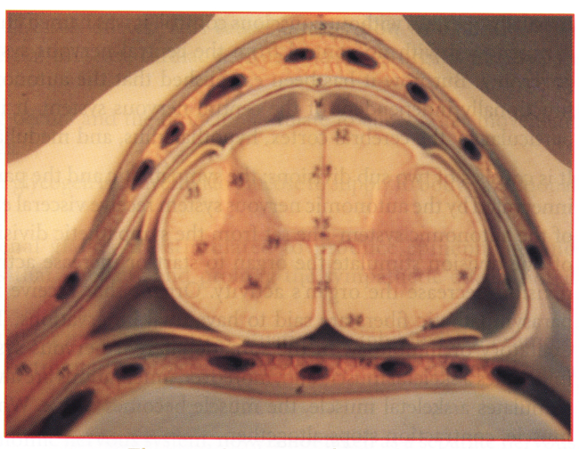
Fig 1-18. Structure of spinal cord.
Functions: The spinal cord has two main functions. First, it is the center for thousands of reflex arcs. The interneuron passes the nerve impulse to a motor neuron that brings about a reflex. The tracts cross over in the medulla, and therefore, the right side of the brain controls the left side of the body, and vice versa. Reflexes allow quick response to a stimulus because they do not require conscious thought.
The spinal cord's second function is that it provides a means of communication between the brain and the peripheral nerves that leave the cord. Sensory impulses travel up the cord to the brain in ascending tracts, and motor impulses travel down the cord from the brain in descending tracts. Sensory impulses that reach the cerebrum result in sensation, and motor impulses that travel from the cerebrum parts. If the spinal cord is injured, we suffer a loss of sensation and a loss of voluntary control-that is, we suffer a paralysis.
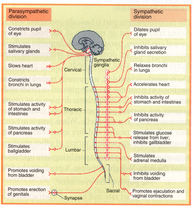
Fig 1-19. The autonomic nervous system.